AmScope B120 Series Binocular Compound Microscope 40X-2500X Magnification with LED, Siedentopf Head, Book and 25 Prepared Slides AmScope B120 Series Binocular Compound Microscope 40X-2500X Magnification with LED, Siedentopf Head, 25 Prepared Slides, Book and 1MP Digital Camera AmScope B120 Series Binocular Compound Microscope 40X-2500X Magnification with LED, Siedentopf Head, 25 Prepared Slides, Book and 1MP Digital Camera AmScope B120 Series Binocular Compound Microscope 40X-2500X Magnification with LED, Siedentopf Head, 25 Prepared Slides, Book and 3MP Digital Camera AmScope B120 Series Binocular Compound Microscope 40X-2500X Magnification with LED, Siedentopf Head, 25 Prepared Slides, Book and 5MP Digital Camera AmScope B120 Series Binocular Compound Microscope 40X-2000X Magnification with LED, Siedentopf Head, 25 Prepared Slides and Book AmScope B120 Series Binocular Compound Microscope 40X-2000X Magnification with LED, Siedentopf Head, 25 Prepared Slides, Book and 1MP Digital Camera AmScope B120 Series Binocular Compound Microscope 40X-2000X Magnification with LED, Siedentopf Head, 25 Prepared Slides, Book and 1MP Digital Camera AmScope B120 Series Binocular Compound Microscope 40X-2000X Magnification with LED, Siedentopf Head, 25 Prepared Slides, Book and 3MP Digital Camera AmScope B120 Series Binocular Compound Microscope 40X-2000X Magnification with LED, Siedentopf Head, 25 Prepared Slides, Book and 5MP Digital Camera
Key Features
- 40X to 2500X expanded magnification range using high-quality, color-coded objective lenses, and 10X and 25X eyepieces for six unique settings.
- An economical microscope for students and professionals, the B120 packs high-quality optics and refined mechanics into a compact form.
- Professional features include a Siedentopf binocular head for precise adjustments, a 2-layer mechanical stage with low-position controls, and coaxial coarse and fine focus for ergonomics and an efficient workflow.
- Includes The World of the Microscope: a beautifully-illustrated book that will help guide first-time users through the history of microscopes and how to perform experiments right at home.
- Includes a set of 25 prepared slides to view on your new microscope, with a variety of plant and animal specimens chosen to provide a range of shapes and colors, ideal for learning to use the microscope, as well as providing a quick study of nature at the microscopic level.
- 40X to 2500X expanded magnification range using high-quality, color-coded objective lenses, and 10X and 25X eyepieces for six unique settings.
- An economical microscope for students and professionals, the B120 packs high-quality optics and refined mechanics into a compact form.
- Professional features include a Siedentopf binocular head for precise adjustments, a 2-layer mechanical stage with low-position controls, and coaxial coarse and fine focus for ergonomics and an efficient workflow.
- Monitor and capture photos and videos with the included 0.3MP digital eyepiece camera. Professional microscopy software for Windows, Mac, and Linux provide tools for image processing, measuring, and more.
- Includes The World of the Microscope: a beautifully-illustrated book that will help guide first-time users through the history of microscopes and how to perform experiments right at home.
- Includes a set of 25 prepared slides to view on your new microscope, with a variety of plant and animal specimens chosen to provide a range of shapes and colors, ideal for learning to use the microscope, as well as providing a quick study of nature at the microscopic level.
- 40X to 2500X expanded magnification range using high-quality, color-coded objective lenses, and 10X and 25X eyepieces for six unique settings.
- An economical microscope for students and professionals, the B120 packs high-quality optics and refined mechanics into a compact form.
- Professional features include a Siedentopf binocular head for precise adjustments, a 2-layer mechanical stage with low-position controls, and coaxial coarse and fine focus for ergonomics and an efficient workflow.
- Monitor and capture photos and videos with the included 1.3MP digital eyepiece camera. Professional microscopy software for Windows, Mac, and Linux provide tools for image processing, measuring, and more.
- Includes The World of the Microscope: a beautifully-illustrated book that will help guide first-time users through the history of microscopes and how to perform experiments right at home.
- Includes a set of 25 prepared slides to view on your new microscope, with a variety of plant and animal specimens chosen to provide a range of shapes and colors, ideal for learning to use the microscope, as well as providing a quick study of nature at the microscopic level.
- 40X to 2500X expanded magnification range using high-quality, color-coded objective lenses, and 10X and 25X eyepieces for six unique settings.
- An economical microscope for students and professionals, the B120 packs high-quality optics and refined mechanics into a compact form.
- Professional features include a Siedentopf binocular head for precise adjustments, a 2-layer mechanical stage with low-position controls, and coaxial coarse and fine focus for ergonomics and an efficient workflow.
- Monitor and capture photos and videos with the included 3MP digital eyepiece camera. Professional microscopy software for Windows, Mac, and Linux provide tools for image processing, measuring, and more.
- Includes The World of the Microscope: a beautifully-illustrated book that will help guide first-time users through the history of microscopes and how to perform experiments right at home.
- Includes a set of 25 prepared slides to view on your new microscope, with a variety of plant and animal specimens chosen to provide a range of shapes and colors, ideal for learning to use the microscope, as well as providing a quick study of nature at the microscopic level.
- 40X to 2500X expanded magnification range using high-quality, color-coded objective lenses, and 10X and 25X eyepieces for six unique settings.
- An economical microscope for students and professionals, the B120 packs high-quality optics and refined mechanics into a compact form.
- Professional features include a Siedentopf binocular head for precise adjustments, a 2-layer mechanical stage with low-position controls, and coaxial coarse and fine focus for ergonomics and an efficient workflow.
- Monitor and capture photos and videos with the included 5MP digital eyepiece camera. Professional microscopy software for Windows, Mac, and Linux provide tools for image processing, measuring, and more.
- Includes The World of the Microscope: a beautifully-illustrated book that will help guide first-time users through the history of microscopes and how to perform experiments right at home.
- Includes a set of 25 prepared slides to view on your new microscope, with a variety of plant and animal specimens chosen to provide a range of shapes and colors, ideal for learning to use the microscope, as well as providing a quick study of nature at the microscopic level.
- 40X to 2000X expanded magnification range using high-quality, color-coded objective lenses, and 10X and 20X eyepieces for eight unique settings.
- An economical microscope for students and professionals, the B120 packs high-quality optics and refined mechanics into a compact form.
- Professional features include a Siedentopf binocular head for precise adjustments, a 2-layer mechanical stage with low-position controls, and coaxial coarse and fine focus for ergonomics and an efficient workflow.
- Monitor and capture photos and videos with the included 0.3MP digital eyepiece camera. Professional microscopy software for Windows, Mac, and Linux provide tools for image processing, measuring, and more.
- Includes The World of the Microscope: a beautifully-illustrated book that will help guide first-time users through the history of microscopes and how to perform experiments right at home.
- Includes a set of 25 prepared slides to view on your new microscope, with a variety of plant and animal specimens chosen to provide a range of shapes and colors, ideal for learning to use the microscope, as well as providing a quick study of nature at the microscopic level.
- Includes an AmScope Exclusive – digital camera software designed for middle and high school students that makes capturing and editing photos and videos easy and fun! Windows and Mac
- 40X to 2000X expanded magnification range using high-quality, color-coded objective lenses, and 10X and 20X eyepieces for eight unique settings.
- An economical microscope for students and professionals, the B120 packs high-quality optics and refined mechanics into a compact form.
- Professional features include a Siedentopf binocular head for precise adjustments, a 2-layer mechanical stage with low-position controls, and coaxial coarse and fine focus for ergonomics and an efficient workflow.
- Monitor and capture photos and videos with the included 1.3MP digital eyepiece camera. Professional microscopy software for Windows, Mac, and Linux provide tools for image processing, measuring, and more.
- Includes The World of the Microscope: a beautifully-illustrated book that will help guide first-time users through the history of microscopes and how to perform experiments right at home.
- Includes a set of 25 prepared slides to view on your new microscope, with a variety of plant and animal specimens chosen to provide a range of shapes and colors, ideal for learning to use the microscope, as well as providing a quick study of nature at the microscopic level.
- Includes an AmScope Exclusive digital camera software designed for middle and high school students that makes capturing and editing photos and videos easy and fun! Windows and Mac
- 40X to 2000X expanded magnification range using high-quality, color-coded objective lenses, and 10X and 20X eyepieces for eight unique settings.
- An economical microscope for students and professionals, the B120 packs high-quality optics and refined mechanics into a compact form.
- Professional features include a Siedentopf binocular head for precise adjustments, a 2-layer mechanical stage with low-position controls, and coaxial coarse and fine focus for ergonomics and an efficient workflow.
- Monitor and capture photos and videos with the included 3MP digital eyepiece camera. Professional microscopy software for Windows, Mac, and Linux provide tools for image processing, measuring, and more.
- Includes The World of the Microscope: a beautifully-illustrated book that will help guide first-time users through the history of microscopes and how to perform experiments right at home.
- Includes a set of 25 prepared slides to view on your new microscope, with a variety of plant and animal specimens chosen to provide a range of shapes and colors, ideal for learning to use the microscope, as well as providing a quick study of nature at the microscopic level.
- Includes an AmScope Exclusive – digital camera software designed for middle and high school students that makes capturing and editing photos and videos easy and fun! Windows and Mac
- 40X to 2000X expanded magnification range using high-quality, color-coded objective lenses, and 10X and 20X eyepieces for eight unique settings.
- An economical microscope for students and professionals, the B120 packs high-quality optics and refined mechanics into a compact form.
- Professional features include a Siedentopf binocular head for precise adjustments, a 2-layer mechanical stage with low-position controls, and coaxial coarse and fine focus for ergonomics and an efficient workflow.
- Monitor and capture photos and videos with the included 5MP digital eyepiece camera. Professional microscopy software for Windows, Mac, and Linux provide tools for image processing, measuring, and more.
- Includes The World of the Microscope: a beautifully-illustrated book that will help guide first-time users through the history of microscopes and how to perform experiments right at home.
- Includes a set of 25 prepared slides to view on your new microscope, with a variety of plant and animal specimens chosen to provide a range of shapes and colors, ideal for learning to use the microscope, as well as providing a quick study of nature at the microscopic level.
- Includes an AmScope Exclusive – digital camera software designed for middle and high school students that makes capturing and editing photos and videos easy and fun! Windows and Mac
The B120 binocular compound microscope is designed for teaching demonstrations, clinical examinations and laboratory applications. It is an ideal microscope for teachers and students, including those in medical school or with a major in biology. It has a number of advancements in functionality compared to other microscopes in its class, such as enhanced LED lighting, centerable condenser, and Siedentopf head adjustments. Use the microscope for studying fixed or live cells, bacteria, plants and soil, or water samples.
Viewing Head

To customize the microscope to your viewing comfort, the eyepiece spacing can be easily adjusted using a precision Siedentopf mechanism. A diopter is available on the left ocular tube to compensate for asymmetry, and the ocular tubes are angled at 30° to comfortably accommodate a seated position without neck strain. Various eyepieces can be used to increase the overall optical magnification.
Optics
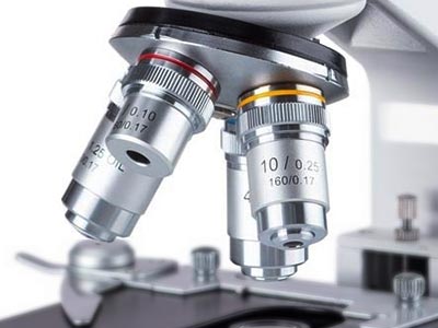
The objective turret provides instant access to 4 magnification levels to easily focus in on minute details from 40X to 1000X. Additional 25X eyepieces expand the maximum magnification to 2500X with 6 unique levels. This covers magnifications needed for studying hair follicles, cells, and bacteria. These high-quality lenses are achromatically corrected to improve resolution and color accuracy.
The Stage
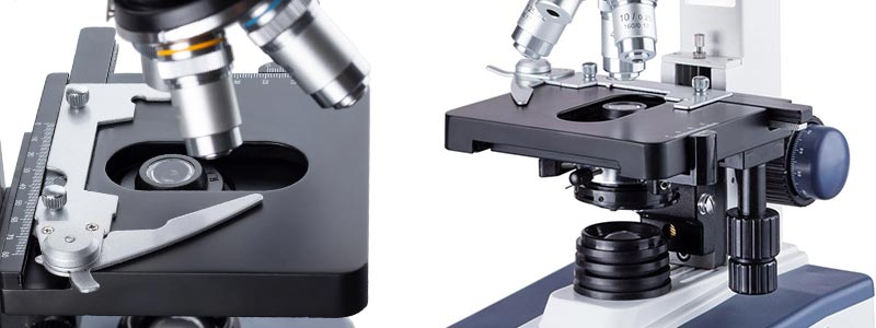
The 2-layer mechanical stage provides smooth and precise movement for examination of specimen slides. The stage's low-position controls are conveniently placed near the coaxial focus knobs for a streamlined workflow. Examining specimen slides is intuitive and precise for users at any skill level.
Transmitted Illumination
The LED sub-stage lighting provides cool, energy-efficient illumination. The light-source is daylight balanced to produce natural colors for imaging. With our specially developed fly-eye lens, the light is evenly distributed at maximum brightness to improve contrast and resolution. Illumination is further enhanced by a centerable Abbe condenser, which can be realigned for any magnification to guarantee proper lighting.
This kit includes a beautifully-illustrated book that will help guide first-time users through the history of microscopes, and how to perform some experiments right at home. The book provides an insight into the history of microscopic study and the invention of microscopes. Several popular experiments are illustrated and explained to guide you through the scientific process. This kit also includes a set of 25 prepared slides to view on your new microscope. A variety of plant and animal specimens are pre-mounted for instant viewing, including cross-sections of plant stems, and cellular structures. These slides are chosen to provide a range of shapes and colors, ideal for learning to use the microscope, as well as providing a quick study of nature at the microscopic level.
Specifications
| Optical System | Finite-conjugate |
| Mechanical Tube Length | 160mm |
| Head | Binocular, 30° incline, 360° rotatable |
| Interpupillary Adjustment | Siedentopf, 53-77mm |
| Ocular Diameter | 23mm |
| Eyepieces | 10X, 25X |
| Objective Lenses | DIN standard |
| Objective Parfocal Distance | 45mm |
| Objective Mounting Thread | RMS 20.32mm |
| Objective Turret | quadruple |
| Focusing System | Coaxial coarse and fine focus |
| Focus Range | 30mm |
| Division of Fine Focus | 0.002mm |
| Stage Design | Double-layer with caliper |
| Stage Dimensions | 118mm x 127mm |
| X-Y Translation Range | 70mm x 21mm |
| Transmitted Illumination | Variable-intensity 1W LED, fly-eye lens |
| Condenser | NA1.25 Abbe condenser with iris diaphragm |
| Sub-stage Condenser-holder | rack and pinion, centerable |
| Power | 220V |
| Dimensions | 11" x 7" x 13-3/4" (28cm x 17.8cm x 35cm) |
Objective Lenses
| Magnification | Corrections | NA | Immersion Medium | Cover-glass Thickness |
|---|---|---|---|---|
| 4X | achromatic | 0.10 | — | 0.17mm |
| 10X | achromatic | 0.25 | — | 0.17mm |
| 40X | achromatic | 0.65 | — | 0.17mm |
| 100X | achromatic | 1.25 | oil | 0.17mm |
Packing List:
The B120 binocular compound microscope is designed for teaching demonstrations, clinical examinations and laboratory applications. It is an ideal microscope for teachers and students, including those in medical school or with a major in biology. It has a number of advancements in functionality compared to other microscopes in its class, such as enhanced LED lighting, centerable condenser, and Siedentopf head adjustments. Use the microscope for studying fixed or live cells, bacteria, plants and soil, or water samples.
Viewing Head
To customize the microscope to your viewing comfort, the eyepiece spacing can be easily adjusted using a precision Siedentopf mechanism. A diopter is available on the left ocular tube to compensate for asymmetry, and the ocular tubes are angled at 30° to comfortably accommodate a seated position without neck strain. Various eyepieces can be used to increase the overall optical magnification.
Optics
The objective turret provides instant access to 4 magnification levels to easily focus in on minute details from 40X to 1000X. Additional 25X eyepieces expand the maximum magnification to 2500X with 6 unique levels. This covers magnifications needed for studying hair follicles, cells, and bacteria. These high-quality lenses are achromatically corrected to improve resolution and color accuracy.
The Stage
The 2-layer mechanical stage provides smooth and precise movement for examination of specimen slides. The stage's low-position controls are conveniently placed near the coaxial focus knobs for a streamlined workflow. Examining specimen slides is intuitive and precise for users at any skill level.
Transmitted Illumination
The LED sub-stage lighting provides cool, energy-efficient illumination. The light-source is daylight balanced to produce natural colors for imaging. With our specially developed fly-eye lens, the light is evenly distributed at maximum brightness to improve contrast and resolution. Illumination is further enhanced by a centerable Abbe condenser, which can be realigned for any magnification to guarantee proper lighting.
Digital Imaging
Monitor and capture photos and videos with the included 1MP digital eyepiece camera. The compact, lightweight camera can be used in place of an eyepiece to watch live images, and to record photos or videos on your computer. This ability to view microscopic images on your computer reduces eye-strain, and allows groups of people to view images at the same time. Our professional software for Windows provides a wide assortment of capture and photo-editing functions including color correction, time-lapse capture, image stitching, and a full complement of measuring tools. A lite version of our software is available for Mac and Linux with essential functionality for capturing photos and videos.
This kit includes a beautifully-illustrated book that will help guide first-time users through the history of microscopes, and how to perform some experiments right at home. The book provides an insight into the history of microscopic study and the invention of microscopes. Several popular experiments are illustrated and explained to guide you through the scientific process. This kit also includes a set of 25 prepared slides to view on your new microscope. A variety of plant and animal specimens are pre-mounted for instant viewing, including cross-sections of plant stems, and cellular structures. These slides are chosen to provide a range of shapes and colors, ideal for learning to use the microscope, as well as providing a quick study of nature at the microscopic level.
Specifications
| Optical System | Finite-conjugate |
| Mechanical Tube Length | 160mm |
| Head | Binocular, 30° incline, 360° rotatable |
| Interpupillary Adjustment | Siedentopf, 53-77mm |
| Ocular Diameter | 23mm |
| Eyepieces | 10X, 25X |
| Objective Lenses | DIN standard |
| Objective Parfocal Distance | 45mm |
| Objective Mounting Thread | RMS 20.32mm |
| Objective Turret | quadruple |
| Focusing System | Coaxial coarse and fine focus |
| Focus Range | 30mm |
| Division of Fine Focus | 0.002mm |
| Stage Design | Double-layer with caliper |
| Stage Dimensions | 118mm x 127mm |
| X-Y Translation Range | 70mm x 21mm |
| Transmitted Illumination | Variable-intensity 1W LED, fly-eye lens |
| Condenser | NA1.25 Abbe condenser with iris diaphragm |
| Sub-stage Condenser-holder | rack and pinion, centerable |
| Power | 220V |
| Dimensions | 11" x 7" x 13-3/4" (28cm x 17.8cm x 35cm) |
Objective Lenses
| Magnification | Corrections | NA | Immersion Medium | Cover-glass Thickness |
|---|---|---|---|---|
| 4X | achromatic | 0.10 | — | 0.17mm |
| 10X | achromatic | 0.25 | — | 0.17mm |
| 40X | achromatic | 0.65 | — | 0.17mm |
| 100X | achromatic | 1.25 | oil | 0.17mm |
| Camera Specifications | |
| Sensor Type | CMOS |
| Sensor Optical Format | 1/3" |
| Active Pixels | 0.3M (640 x 480) |
| Pixel Size | 5.6µm x 5.6µm |
| Active Sensor Area | 3.58mm x 2.69mm |
| Shutter | electronic rolling shutter |
| Sensitivity | 13.5V/Lux-sec |
| Spectral Response | 380-650nm with IR-cut filter |
| Capture Resolution and Maximum Framerate | 25fps @ 640x480 |
| Connectivity | USB 2.0 |
| Power | 5VDC over USB |
Software
| OS Requirements | Windows (32/64 bit) XP/Vista/7/8/10, Mac OS 10.8+, Linux kernel 3.13+ | |||||||||||||||
|---|---|---|---|---|---|---|---|---|---|---|---|---|---|---|---|---|
| Hardware Requirements | Intel Core2 2.8GHz or comparable processor, 4GB RAM | |||||||||||||||
| Features |
|
Packing List:
Our binocular compound microscope with a 1.3MP USB imager (camera) is designed for teaching demonstrations, clinical examinations and laboratory applications. It is a perfect microscope for a range of people, from students in high school to teachers including those with a major in biology, or enrolled in medical school. It comes with a professional Siedentopf binocular head, double layer mechanical stage, course & fine focusing, LED illumination, and a 1.3MP USB digital imager.
The digital camera captures still images and video, and allows you to view a live stream on your PC. The included software for Windows offers image-development and measurement tools, as well as advanced compositing features such as image-stitching and extended-depth-of-focus. Independent preview and capture resolutions allow you to view high-speed video while capturing high-definition images.
This microscope is an ideal instrument for bacterial, biological, and pharmaceutical researches.
Microscope Features
- 30 degree inclined 360-degree swiveling Siedentopf binocular head
- High quality precise optical glass elements
- Four Achromatic objectives, DIN 4X, 10X, 40X(S), 100X(S,Oil)
- Two pairs of widefield eyepieces: 10X and 25X
- Adjustable interpupillary distance
- Adjustable ocular diopter
- Variable intensity illumination
- Coaxial coarse and fine focus adjustment
- Focusing knobs are on both sides of the microscope
- Stage Upward Moving Lock protects objectives and slides
- Stain-resistant double layer mechanical stage
- NA1.25 Abbe condenser with iris diaphragm & filters
- Rack and pinion adjustment condenser
Specifications
- Eyepieces: 10X and 25X
- Objectives: Achromatic DIN 4X, 10X, 40X(S), 100X(S, Oil)
- Head: 30 degree inclined 360 degree swiveling binocular
- Interpupillary distance: 2-1/8" - 3-1/8" (53mm - 77mm)
- Adjustable ocular diopter
- Nosepiece: revolving quadruple
- Mechanical Stage: 3-D double layer
- -Size: 4-5/8" x 5" (118mm x 127mm)
- - Travel Range (X-Y): 70mm x 21mm
- Condenser: NA1.25 Abbe condenser with iris diaphragm
- Illumination: LED light, transmitted, intensity adjustable, GS and CE approved
- Focusing: coaxial coarse & fine knobs on both sides
- Power Supply: 100-240VAC, 220V UK Plug w/ EU Adapter (UL approved)
- Dimension: 11" x 7" x 13-3/4" (280mm x 178mm x 350mm)
Camera Specifications
- Sensor: Aptina
- Sensor Type: CMOS
- Sensor Size: 4.6x3.7mm
- Pixel Size: 3.6
- Resolution: 1.3MP
- Frame Rate: 7.5@1280x1024,12.5@1024x768,12.5@800x600
- Compatibility: Windows (32/64 bit) XP/Vista/7/8/10, Mac OSX (Mac software not provided)
Software Specifications
- OS Requirements: Windows (32/64 bit) XP/Vista/7/8/10
- Hardware Requirements: Intel Core2 2.8GHz or comparable processor, 2GB RAM, USB2.0 port
Packing List
- One Siedentopf Binocular Microscope Head
- One Microscope Body Frame with Double Layer Mechanical Stage and LED Illumination
- Four DIN Standard Objectives, 4X, 10X, 40X and 100X
- One Pair of Widefield 10X Eyepieces
- One Pair of Widefield 25X Eyepieces
- One Blue Color Filter
- One Dust Cover
- Sample Immersion Oil
- One 1.3MP USB2.0 Digital Imager
One 6' (1.8m) USB Cable
The B120 binocular compound microscope is designed for teaching demonstrations, clinical examinations and laboratory applications. It is an ideal microscope for teachers and students, including those in medical school or with a major in biology. It has a number of advancements in functionality compared to other microscopes in its class, such as enhanced LED lighting, centerable condenser, and Siedentopf head adjustments. Use the microscope for studying fixed or live cells, bacteria, plants and soil, or water samples.
Viewing Head
To customize the microscope to your viewing comfort, the eyepiece spacing can be easily adjusted using a precision Siedentopf mechanism. A diopter is available on the left ocular tube to compensate for asymmetry, and the ocular tubes are angled at 30° to comfortably accommodate a seated position without neck strain. Various eyepieces can be used to increase the overall optical magnification.
Optics
The objective turret provides instant access to 4 magnification levels to easily focus in on minute details from 40X to 1000X. Additional 25X eyepieces expand the maximum magnification to 2500X with 6 unique levels. This covers magnifications needed for studying hair follicles, cells, and bacteria. These high-quality lenses are achromatically corrected to improve resolution and color accuracy.
The Stage
The 2-layer mechanical stage provides smooth and precise movement for examination of specimen slides. The stage's low-position controls are conveniently placed near the coaxial focus knobs for a streamlined workflow. Examining specimen slides is intuitive and precise for users at any skill level.
Transmitted Illumination
The LED sub-stage lighting provides cool, energy-efficient illumination. The light-source is daylight balanced to produce natural colors for imaging. With our specially developed fly-eye lens, the light is evenly distributed at maximum brightness to improve contrast and resolution. Illumination is further enhanced by a centerable Abbe condenser, which can be realigned for any magnification to guarantee proper lighting.
Digital Imaging
Monitor and capture photos and videos with the included 3MP digital eyepiece camera. The compact, lightweight camera can be used in place of an eyepiece to watch live images, and to record photos or videos on your computer. This ability to view microscopic images on your computer reduces eye-strain, and allows groups of people to view images at the same time. Our professional software for Windows provides a wide assortment of capture and photo-editing functions including color correction, time-lapse capture, image stitching, and a full complement of measuring tools. A lite version of our software is available for Mac and Linux with essential functionality for capturing photos and videos.
This kit includes a beautifully-illustrated book that will help guide first-time users through the history of microscopes, and how to perform some experiments right at home. The book provides an insight into the history of microscopic study and the invention of microscopes. Several popular experiments are illustrated and explained to guide you through the scientific process. This kit also includes a set of 25 prepared slides to view on your new microscope. A variety of plant and animal specimens are pre-mounted for instant viewing, including cross-sections of plant stems, and cellular structures. These slides are chosen to provide a range of shapes and colors, ideal for learning to use the microscope, as well as providing a quick study of nature at the microscopic level.
Specifications
| Optical System | Finite-conjugate |
| Mechanical Tube Length | 160mm |
| Head | Binocular, 30° incline, 360° rotatable |
| Interpupillary Adjustment | Siedentopf, 53-77mm |
| Ocular Diameter | 23mm |
| Eyepieces | 10X, 25X |
| Objective Lenses | DIN standard |
| Objective Parfocal Distance | 45mm |
| Objective Mounting Thread | RMS 20.32mm |
| Objective Turret | quadruple |
| Focusing System | Coaxial coarse and fine focus |
| Focus Range | 30mm |
| Division of Fine Focus | 0.002mm |
| Stage Design | Double-layer with caliper |
| Stage Dimensions | 118mm x 127mm |
| X-Y Translation Range | 70mm x 21mm |
| Transmitted Illumination | Variable-intensity 1W LED, fly-eye lens |
| Condenser | NA1.25 Abbe condenser with iris diaphragm |
| Sub-stage Condenser-holder | rack and pinion, centerable |
| Power | 220V |
| Dimensions | 11" x 7" x 13-3/4" (28cm x 17.8cm x 35cm) |
Objective Lenses
| Magnification | Corrections | NA | Immersion Medium | Cover-glass Thickness |
|---|---|---|---|---|
| 4X | achromatic | 0.10 | — | 0.17mm |
| 10X | achromatic | 0.25 | — | 0.17mm |
| 40X | achromatic | 0.65 | — | 0.17mm |
| 100X | achromatic | 1.25 | oil | 0.17mm |
| Camera Specifications | |
| Sensor Type | CMOS |
| Sensor Optical Format | 1/2.7" |
| Active Pixels | 3M (2048 x 1536) |
| Pixel Size | 2.2µm x 2.2µm |
| Active Sensor Area | 4.51mm x 3.38mm |
| Shutter | electronic rolling shutter |
| Spectral Response | 380-650nm with IR-cut filter |
| Capture Resolution and Maximum Framerate | 3fps @ 2048x1536 5fps @ 1600x1200 7.5fps @ 1280x1024 |
| Connectivity | USB 2.0 |
| Power | 5VDC over USB |
Software
| OS Requirements | Windows (32/64 bit) XP/Vista/7/8/10, Mac OS 10.8+, Linux kernel 3.13+ | |||||||||||||||
|---|---|---|---|---|---|---|---|---|---|---|---|---|---|---|---|---|
| Hardware Requirements | Intel Core2 2.8GHz or comparable processor, 4GB RAM | |||||||||||||||
| Features |
|
Packing List:
The B120 binocular compound microscope is designed for teaching demonstrations, clinical examinations and laboratory applications. It is an ideal microscope for teachers and students, including those in medical school or with a major in biology. It has a number of advancements in functionality compared to other microscopes in its class, such as enhanced LED lighting, centerable condenser, and Siedentopf head adjustments. Use the microscope for studying fixed or live cells, bacteria, plants and soil, or water samples.
Viewing Head
To customize the microscope to your viewing comfort, the eyepiece spacing can be easily adjusted using a precision Siedentopf mechanism. A diopter is available on the left ocular tube to compensate for asymmetry, and the ocular tubes are angled at 30° to comfortably accommodate a seated position without neck strain. Various eyepieces can be used to increase the overall optical magnification.
Optics
The objective turret provides instant access to 4 magnification levels to easily focus in on minute details from 40X to 1000X. Additional 25X eyepieces expand the maximum magnification to 2500X with 6 unique levels. This covers magnifications needed for studying hair follicles, cells, and bacteria. These high-quality lenses are achromatically corrected to improve resolution and color accuracy.
The Stage
The 2-layer mechanical stage provides smooth and precise movement for examination of specimen slides. The stage's low-position controls are conveniently placed near the coaxial focus knobs for a streamlined workflow. Examining specimen slides is intuitive and precise for users at any skill level.
Transmitted Illumination
The LED sub-stage lighting provides cool, energy-efficient illumination. The light-source is daylight balanced to produce natural colors for imaging. With our specially developed fly-eye lens, the light is evenly distributed at maximum brightness to improve contrast and resolution. Illumination is further enhanced by a centerable Abbe condenser, which can be realigned for any magnification to guarantee proper lighting.
Digital Imaging
Monitor and capture photos and videos with the included 5MP digital eyepiece camera. The compact, lightweight camera can be used in place of an eyepiece to watch live images, and to record photos or videos on your computer. This ability to view microscopic images on your computer reduces eye-strain, and allows groups of people to view images at the same time. Our professional software for Windows provides a wide assortment of capture and photo-editing functions including color correction, time-lapse capture, image stitching, and a full complement of measuring tools. A lite version of our software is available for Mac and Linux with essential functionality for capturing photos and videos.
This kit includes a beautifully-illustrated book that will help guide first-time users through the history of microscopes, and how to perform some experiments right at home. The book provides an insight into the history of microscopic study and the invention of microscopes. Several popular experiments are illustrated and explained to guide you through the scientific process. This kit also includes a set of 25 prepared slides to view on your new microscope. A variety of plant and animal specimens are pre-mounted for instant viewing, including cross-sections of plant stems, and cellular structures. These slides are chosen to provide a range of shapes and colors, ideal for learning to use the microscope, as well as providing a quick study of nature at the microscopic level.
Specifications
| Optical System | Finite-conjugate |
| Mechanical Tube Length | 160mm |
| Head | Binocular, 30° incline, 360° rotatable |
| Interpupillary Adjustment | Siedentopf, 53-77mm |
| Ocular Diameter | 23mm |
| Eyepieces | 10X, 25X |
| Objective Lenses | DIN standard |
| Objective Parfocal Distance | 45mm |
| Objective Mounting Thread | RMS 20.32mm |
| Objective Turret | quadruple |
| Focusing System | Coaxial coarse and fine focus |
| Focus Range | 30mm |
| Division of Fine Focus | 0.002mm |
| Stage Design | Double-layer with caliper |
| Stage Dimensions | 118mm x 127mm |
| X-Y Translation Range | 70mm x 21mm |
| Transmitted Illumination | Variable-intensity 1W LED, fly-eye lens |
| Condenser | NA1.25 Abbe condenser with iris diaphragm |
| Sub-stage Condenser-holder | rack and pinion, centerable |
| Power | 220V |
| Dimensions | 11" x 7" x 13-3/4" (28cm x 17.8cm x 35cm) |
Objective Lenses
| Magnification | Corrections | NA | Immersion Medium | Cover-glass Thickness |
|---|---|---|---|---|
| 4X | achromatic | 0.10 | — | 0.17mm |
| 10X | achromatic | 0.25 | — | 0.17mm |
| 40X | achromatic | 0.65 | — | 0.17mm |
| 100X | achromatic | 1.25 | oil | 0.17mm |
| Camera Specifications | |
| Sensor Type | CMOS |
| Sensor Optical Format | 1/2.5" |
| Active Pixels | 5M (2592 x 1944) |
| Pixel Size | 2.2µm x 2.2µm |
| Active Sensor Area | 5.7mm x 4.28mm |
| Shutter | electronic rolling shutter |
| Spectral Response | 380-650nm with IR-cut filter |
| Capture Resolution and Maximum Framerate | 2fps @ 2592x1944 5fps @ 1600x1200 7.5fps @ 1280x1024 |
| Connectivity | USB 2.0 |
| Power | 5VDC over USB |
Software
| OS Requirements | Windows (32/64 bit) XP/Vista/7/8/10, Mac OS 10.8+, Linux kernel 3.13+ | |||||||||||||||
|---|---|---|---|---|---|---|---|---|---|---|---|---|---|---|---|---|
| Hardware Requirements | Intel Core2 2.8GHz or comparable processor, 4GB RAM | |||||||||||||||
| Features |
|
Packing List:
The B120 binocular compound microscope is designed for teaching demonstrations, clinical examinations and laboratory applications. It is an ideal microscope for teachers and students, including those in medical school or with a major in biology. It has a number of advancements in functionality compared to other microscopes in its class, such as enhanced LED lighting, centerable condenser, and Siedentopf head adjustments. Use the microscope for studying fixed or live cells, bacteria, plants and soil, or water samples.
Viewing Head
To customize the microscope to your viewing comfort, the eyepiece spacing can be easily adjusted using a precision Siedentopf mechanism. A diopter is available on the left ocular tube to compensate for asymmetry, and the ocular tubes are angled at 30° to comfortably accommodate a seated position without neck strain. Various eyepieces can be used to increase the overall optical magnification.
Optics
The objective turret provides instant access to 4 magnification levels to easily focus in on minute details from 40X to 1000X. Additional 20X eyepieces expand the maximum magnification to 2000X with 8 unique levels. This covers magnifications needed for studying hair follicles, cells, and bacteria. These high-quality lenses are achromatically corrected to improve resolution and color accuracy.
The Stage
The 2-layer mechanical stage provides smooth and precise movement for examination of specimen slides. The stage's low-position controls are conveniently placed near the coaxial focus knobs for a streamlined workflow. Examining specimen slides is intuitive and precise for users at any skill level.
Transmitted Illumination
The LED sub-stage lighting provides cool, energy-efficient illumination. The light-source is daylight balanced to produce natural colors for imaging. With our specially developed fly-eye lens, the light is evenly distributed at maximum brightness to improve contrast and resolution. Illumination is further enhanced by a centerable Abbe condenser, which can be realigned for any magnification to guarantee proper lighting.
This kit includes a beautifully-illustrated book that will help guide first-time users through the history of microscopes, and how to perform some experiments right at home. The book provides an insight into the history of microscopic study and the invention of microscopes. Several popular experiments are illustrated and explained to guide you through the scientific process. This kit also includes a set of 25 prepared slides to view on your new microscope. A variety of plant and animal specimens are pre-mounted for instant viewing, including cross-sections of plant stems, and cellular structures. These slides are chosen to provide a range of shapes and colors, ideal for learning to use the microscope, as well as providing a quick study of nature at the microscopic level.
Specifications
| Optical System | Finite-conjugate |
| Mechanical Tube Length | 160mm |
| Head | Binocular, 30° incline, 360° rotatable |
| Interpupillary Adjustment | Siedentopf, 53-77mm |
| Ocular Diameter | 23mm |
| Eyepieces | 10X, 20X |
| Objective Lenses | DIN standard |
| Objective Parfocal Distance | 45mm |
| Objective Mounting Thread | RMS 20.32mm |
| Objective Turret | quadruple |
| Focusing System | Coaxial coarse and fine focus |
| Focus Range | 30mm |
| Division of Fine Focus | 0.002mm |
| Stage Design | Double-layer with caliper |
| Stage Dimensions | 118mm x 127mm |
| X-Y Translation Range | 70mm x 21mm |
| Transmitted Illumination | Variable-intensity 1W LED, fly-eye lens |
| Condenser | NA1.25 Abbe condenser with iris diaphragm |
| Sub-stage Condenser-holder | rack and pinion, centerable |
| Power | 100-240VAC 50/60Hz wide-band |
| Dimensions | 11" x 7" x 13-3/4" (28cm x 17.8cm x 35cm) |
Objective Lenses
| Magnification | Corrections | NA | Immersion Medium | Cover-glass Thickness |
|---|---|---|---|---|
| 4X | achromatic | 0.10 | — | 0.17mm |
| 10X | achromatic | 0.25 | — | 0.17mm |
| 40X | achromatic | 0.65 | — | 0.17mm |
| 100X | achromatic | 1.25 | oil | 0.17mm |
Packing List:
The B120 binocular compound microscope is designed for teaching demonstrations, clinical examinations and laboratory applications. It is an ideal microscope for teachers and students, including those in medical school or with a major in biology. It has a number of advancements in functionality compared to other microscopes in its class, such as enhanced LED lighting, centerable condenser, and Siedentopf head adjustments. Use the microscope for studying fixed or live cells, bacteria, plants and soil, or water samples.
Viewing Head
To customize the microscope to your viewing comfort, the eyepiece spacing can be easily adjusted using a precision Siedentopf mechanism. A diopter is available on the left ocular tube to compensate for asymmetry, and the ocular tubes are angled at 30° to comfortably accommodate a seated position without neck strain. Various eyepieces can be used to increase the overall optical magnification.
Optics
The objective turret provides instant access to 4 magnification levels to easily focus in on minute details from 40X to 1000X. Additional 20X eyepieces expand the maximum magnification to 2000X with 8 unique levels. This covers magnifications needed for studying hair follicles, cells, and bacteria. These high-quality lenses are achromatically corrected to improve resolution and color accuracy.
The Stage
The 2-layer mechanical stage provides smooth and precise movement for examination of specimen slides. The stage's low-position controls are conveniently placed near the coaxial focus knobs for a streamlined workflow. Examining specimen slides is intuitive and precise for users at any skill level.
Transmitted Illumination
The LED sub-stage lighting provides cool, energy-efficient illumination. The light-source is daylight balanced to produce natural colors for imaging. With our specially developed fly-eye lens, the light is evenly distributed at maximum brightness to improve contrast and resolution. Illumination is further enhanced by a centerable Abbe condenser, which can be realigned for any magnification to guarantee proper lighting.
Digital Imaging
Monitor and capture photos and videos with the included 0.3MP digital eyepiece camera. The compact, lightweight camera can be used in place of an eyepiece to watch live images, and to record photos or videos on your computer. This ability to view microscopic images on your computer reduces eye-strain, and allows groups of people to view images at the same time. Our professional software for Windows provides a wide assortment of capture and photo-editing functions including color correction, time-lapse capture, image stitching, and a full complement of measuring tools. A lite version of our software is available for Mac and Linux with essential functionality for capturing photos and videos.
This kit includes a beautifully-illustrated book that will help guide first-time users through the history of microscopes, and how to perform some experiments right at home. The book provides an insight into the history of microscopic study and the invention of microscopes. Several popular experiments are illustrated and explained to guide you through the scientific process. This kit also includes a set of 25 prepared slides to view on your new microscope. A variety of plant and animal specimens are pre-mounted for instant viewing, including cross-sections of plant stems, and cellular structures. These slides are chosen to provide a range of shapes and colors, ideal for learning to use the microscope, as well as providing a quick study of nature at the microscopic level.
Specifications
| Optical System | Finite-conjugate |
| Mechanical Tube Length | 160mm |
| Head | Binocular, 30° incline, 360° rotatable |
| Interpupillary Adjustment | Siedentopf, 53-77mm |
| Ocular Diameter | 23mm |
| Eyepieces | 10X, 20X |
| Objective Lenses | DIN standard |
| Objective Parfocal Distance | 45mm |
| Objective Mounting Thread | RMS 20.32mm |
| Objective Turret | quadruple |
| Focusing System | Coaxial coarse and fine focus |
| Focus Range | 30mm |
| Division of Fine Focus | 0.002mm |
| Stage Design | Double-layer with caliper |
| Stage Dimensions | 118mm x 127mm |
| X-Y Translation Range | 70mm x 21mm |
| Transmitted Illumination | Variable-intensity 1W LED, fly-eye lens |
| Condenser | NA1.25 Abbe condenser with iris diaphragm |
| Sub-stage Condenser-holder | rack and pinion, centerable |
| Power | 100-240VAC 50/60Hz wide-band |
| Dimensions | 11" x 7" x 13-3/4" (28cm x 17.8cm x 35cm) |
Objective Lenses
| Magnification | Corrections | NA | Immersion Medium | Cover-glass Thickness |
|---|---|---|---|---|
| 4X | achromatic | 0.10 | — | 0.17mm |
| 10X | achromatic | 0.25 | — | 0.17mm |
| 40X | achromatic | 0.65 | — | 0.17mm |
| 100X | achromatic | 1.25 | oil | 0.17mm |
| Camera Specifications | |
| Sensor Type | CMOS |
| Sensor Optical Format | 1/3" |
| Active Pixels | 0.3M (640 x 480) |
| Pixel Size | 5.6µm x 5.6µm |
| Active Sensor Area | 3.58mm x 2.69mm |
| Shutter | electronic rolling shutter |
| Sensitivity | 13.5V/Lux-sec |
| Spectral Response | 380-650nm with IR-cut filter |
| Capture Resolution and Maximum Framerate | 25fps @ 640x480 |
| Connectivity | USB 2.0 |
| Power | 5VDC over USB |
Software
| OS Requirements | Windows (32/64 bit) XP/Vista/7/8/10, Mac OS 10.8+, Linux kernel 3.13+ | |||||||||||||||
|---|---|---|---|---|---|---|---|---|---|---|---|---|---|---|---|---|
| Hardware Requirements | Intel Core2 2.8GHz or comparable processor, 4GB RAM | |||||||||||||||
| Features |
|
Packing List:
The B120 binocular compound microscope is designed for teaching demonstrations, clinical examinations and laboratory applications. It is an ideal microscope for teachers and students, including those in medical school or with a major in biology. It has a number of advancements in functionality compared to other microscopes in its class, such as enhanced LED lighting, centerable condenser, and Siedentopf head adjustments. Use the microscope for studying fixed or live cells, bacteria, plants and soil, or water samples.
Viewing Head
To customize the microscope to your viewing comfort, the eyepiece spacing can be easily adjusted using a precision Siedentopf mechanism. A diopter is available on the left ocular tube to compensate for asymmetry, and the ocular tubes are angled at 30° to comfortably accommodate a seated position without neck strain. Various eyepieces can be used to increase the overall optical magnification.
Optics
The objective turret provides instant access to 4 magnification levels to easily focus in on minute details from 40X to 1000X. Additional 20X eyepieces expand the maximum magnification to 2000X with 8 unique levels. This covers magnifications needed for studying hair follicles, cells, and bacteria. These high-quality lenses are achromatically corrected to improve resolution and color accuracy.
The Stage
The 2-layer mechanical stage provides smooth and precise movement for examination of specimen slides. The stage's low-position controls are conveniently placed near the coaxial focus knobs for a streamlined workflow. Examining specimen slides is intuitive and precise for users at any skill level.
Transmitted Illumination
The LED sub-stage lighting provides cool, energy-efficient illumination. The light-source is daylight balanced to produce natural colors for imaging. With our specially developed fly-eye lens, the light is evenly distributed at maximum brightness to improve contrast and resolution. Illumination is further enhanced by a centerable Abbe condenser, which can be realigned for any magnification to guarantee proper lighting.
Digital Imaging
Monitor and capture photos and videos with the included 1.3MP digital eyepiece camera. The compact, lightweight camera can be used in place of an eyepiece to watch live images, and to record photos or videos on your computer. This ability to view microscopic images on your computer reduces eye-strain, and allows groups of people to view images at the same time. Our professional software for Windows provides a wide assortment of capture and photo-editing functions including color correction, time-lapse capture, image stitching, and a full complement of measuring tools. A lite version of our software is available for Mac and Linux with essential functionality for capturing photos and videos.
This kit includes a beautifully-illustrated book that will help guide first-time users through the history of microscopes, and how to perform some experiments right at home. The book provides an insight into the history of microscopic study and the invention of microscopes. Several popular experiments are illustrated and explained to guide you through the scientific process. This kit also includes a set of 25 prepared slides to view on your new microscope. A variety of plant and animal specimens are pre-mounted for instant viewing, including cross-sections of plant stems, and cellular structures. These slides are chosen to provide a range of shapes and colors, ideal for learning to use the microscope, as well as providing a quick study of nature at the microscopic level.
Specifications
| Optical System | Finite-conjugate |
| Mechanical Tube Length | 160mm |
| Head | Binocular, 30° incline, 360° rotatable |
| Interpupillary Adjustment | Siedentopf, 53-77mm |
| Ocular Diameter | 23mm |
| Eyepieces | 10X, 20X |
| Objective Lenses | DIN standard |
| Objective Parfocal Distance | 45mm |
| Objective Mounting Thread | RMS 20.32mm |
| Objective Turret | quadruple |
| Focusing System | Coaxial coarse and fine focus |
| Focus Range | 30mm |
| Division of Fine Focus | 0.002mm |
| Stage Design | Double-layer with caliper |
| Stage Dimensions | 118mm x 127mm |
| X-Y Translation Range | 70mm x 21mm |
| Transmitted Illumination | Variable-intensity 1W LED, fly-eye lens |
| Condenser | NA1.25 Abbe condenser with iris diaphragm |
| Sub-stage Condenser-holder | rack and pinion, centerable |
| Power | 100-240VAC 50/60Hz wide-band |
| Dimensions | 11" x 7" x 13-3/4" (28cm x 17.8cm x 35cm) |
Objective Lenses
| Magnification | Corrections | NA | Immersion Medium | Cover-glass Thickness |
|---|---|---|---|---|
| 4X | achromatic | 0.10 | 0.17mm | |
| 10X | achromatic | 0.25 | 0.17mm | |
| 40X | achromatic | 0.65 | 0.17mm | |
| 100X | achromatic | 1.25 | oil | 0.17mm |
| Camera Specifications | |
| Sensor Type | CMOS |
| Sensor Optical Format | 1/3" |
| Active Pixels | 1M (1280 x 1024) |
| Pixel Size | 3.6µm x 3.6µm |
| Active Sensor Area | 4.61mm x 3.69mm |
| Shutter | electronic rolling shutter |
| Spectral Response | 380-650nm with IR-cut filter |
| Capture Resolution and Maximum Framerate | 7.5fps @ 1280x1024 12.5fps @ 1024x768 12.5fps @ 800x600 |
| Connectivity | USB 2.0 |
| Power | 5VDC over USB |
Software
| OS Requirements | Windows (32/64 bit) XP/Vista/7/8/10, Mac OS 10.8+, Linux kernel 3.13+ | |||||||||||||||
|---|---|---|---|---|---|---|---|---|---|---|---|---|---|---|---|---|
| Hardware Requirements | Intel Core2 2.8GHz or comparable processor, 4GB RAM | |||||||||||||||
| Features |
|
Packing List:
The B120 binocular compound microscope is designed for teaching demonstrations, clinical examinations and laboratory applications. It is an ideal microscope for teachers and students, including those in medical school or with a major in biology. It has a number of advancements in functionality compared to other microscopes in its class, such as enhanced LED lighting, centerable condenser, and Siedentopf head adjustments. Use the microscope for studying fixed or live cells, bacteria, plants and soil, or water samples.
Viewing Head
To customize the microscope to your viewing comfort, the eyepiece spacing can be easily adjusted using a precision Siedentopf mechanism. A diopter is available on the left ocular tube to compensate for asymmetry, and the ocular tubes are angled at 30° to comfortably accommodate a seated position without neck strain. Various eyepieces can be used to increase the overall optical magnification.
Optics
The objective turret provides instant access to 4 magnification levels to easily focus in on minute details from 40X to 1000X. Additional 20X eyepieces expand the maximum magnification to 2000X with 8 unique levels. This covers magnifications needed for studying hair follicles, cells, and bacteria. These high-quality lenses are achromatically corrected to improve resolution and color accuracy.
The Stage
The 2-layer mechanical stage provides smooth and precise movement for examination of specimen slides. The stage's low-position controls are conveniently placed near the coaxial focus knobs for a streamlined workflow. Examining specimen slides is intuitive and precise for users at any skill level.
Transmitted Illumination
The LED sub-stage lighting provides cool, energy-efficient illumination. The light-source is daylight balanced to produce natural colors for imaging. With our specially developed fly-eye lens, the light is evenly distributed at maximum brightness to improve contrast and resolution. Illumination is further enhanced by a centerable Abbe condenser, which can be realigned for any magnification to guarantee proper lighting.
Digital Imaging
Monitor and capture photos and videos with the included 3MP digital eyepiece camera. The compact, lightweight camera can be used in place of an eyepiece to watch live images, and to record photos or videos on your computer. This ability to view microscopic images on your computer reduces eye-strain, and allows groups of people to view images at the same time. Our professional software for Windows provides a wide assortment of capture and photo-editing functions including color correction, time-lapse capture, image stitching, and a full complement of measuring tools. A lite version of our software is available for Mac and Linux with essential functionality for capturing photos and videos.
This kit includes a beautifully-illustrated book that will help guide first-time users through the history of microscopes, and how to perform some experiments right at home. The book provides an insight into the history of microscopic study and the invention of microscopes. Several popular experiments are illustrated and explained to guide you through the scientific process. This kit also includes a set of 25 prepared slides to view on your new microscope. A variety of plant and animal specimens are pre-mounted for instant viewing, including cross-sections of plant stems, and cellular structures. These slides are chosen to provide a range of shapes and colors, ideal for learning to use the microscope, as well as providing a quick study of nature at the microscopic level.
Specifications
| Optical System | Finite-conjugate |
| Mechanical Tube Length | 160mm |
| Head | Binocular, 30° incline, 360° rotatable |
| Interpupillary Adjustment | Siedentopf, 53-77mm |
| Ocular Diameter | 23mm |
| Eyepieces | 10X, 20X |
| Objective Lenses | DIN standard |
| Objective Parfocal Distance | 45mm |
| Objective Mounting Thread | RMS 20.32mm |
| Objective Turret | quadruple |
| Focusing System | Coaxial coarse and fine focus |
| Focus Range | 30mm |
| Division of Fine Focus | 0.002mm |
| Stage Design | Double-layer with caliper |
| Stage Dimensions | 118mm x 127mm |
| X-Y Translation Range | 70mm x 21mm |
| Transmitted Illumination | Variable-intensity 1W LED, fly-eye lens |
| Condenser | NA1.25 Abbe condenser with iris diaphragm |
| Sub-stage Condenser-holder | rack and pinion, centerable |
| Power | 100-240VAC 50/60Hz wide-band |
| Dimensions | 11" x 7" x 13-3/4" (28cm x 17.8cm x 35cm) |
Objective Lenses
| Magnification | Corrections | NA | Immersion Medium | Cover-glass Thickness |
|---|---|---|---|---|
| 4X | achromatic | 0.10 | — | 0.17mm |
| 10X | achromatic | 0.25 | — | 0.17mm |
| 40X | achromatic | 0.65 | — | 0.17mm |
| 100X | achromatic | 1.25 | oil | 0.17mm |
| Camera Specifications | |
| Sensor Type | CMOS |
| Sensor Optical Format | 1/2.7" |
| Active Pixels | 3M (2048 x 1536) |
| Pixel Size | 2.2µm x 2.2µm |
| Active Sensor Area | 4.51mm x 3.38mm |
| Shutter | electronic rolling shutter |
| Spectral Response | 380-650nm with IR-cut filter |
| Capture Resolution and Maximum Framerate | 3fps @ 2048x1536 5fps @ 1600x1200 7.5fps @ 1280x1024 |
| Connectivity | USB 2.0 |
| Power | 5VDC over USB |
Software
| OS Requirements | Windows (32/64 bit) XP/Vista/7/8/10, Mac OS 10.8+, Linux kernel 3.13+ | |||||||||||||||
|---|---|---|---|---|---|---|---|---|---|---|---|---|---|---|---|---|
| Hardware Requirements | Intel Core2 2.8GHz or comparable processor, 4GB RAM | |||||||||||||||
| Features |
|
Packing List:
The B120 binocular compound microscope is designed for teaching demonstrations, clinical examinations and laboratory applications. It is an ideal microscope for teachers and students, including those in medical school or with a major in biology. It has a number of advancements in functionality compared to other microscopes in its class, such as enhanced LED lighting, centerable condenser, and Siedentopf head adjustments. Use the microscope for studying fixed or live cells, bacteria, plants and soil, or water samples.
Viewing Head
To customize the microscope to your viewing comfort, the eyepiece spacing can be easily adjusted using a precision Siedentopf mechanism. A diopter is available on the left ocular tube to compensate for asymmetry, and the ocular tubes are angled at 30° to comfortably accommodate a seated position without neck strain. Various eyepieces can be used to increase the overall optical magnification.
Optics
The objective turret provides instant access to 4 magnification levels to easily focus in on minute details from 40X to 1000X. Additional 20X eyepieces expand the maximum magnification to 2000X with 8 unique levels. This covers magnifications needed for studying hair follicles, cells, and bacteria. These high-quality lenses are achromatically corrected to improve resolution and color accuracy.
The Stage
The 2-layer mechanical stage provides smooth and precise movement for examination of specimen slides. The stage's low-position controls are conveniently placed near the coaxial focus knobs for a streamlined workflow. Examining specimen slides is intuitive and precise for users at any skill level.
Transmitted Illumination
The LED sub-stage lighting provides cool, energy-efficient illumination. The light-source is daylight balanced to produce natural colors for imaging. With our specially developed fly-eye lens, the light is evenly distributed at maximum brightness to improve contrast and resolution. Illumination is further enhanced by a centerable Abbe condenser, which can be realigned for any magnification to guarantee proper lighting.
Digital Imaging
Monitor and capture photos and videos with the included 5MP digital eyepiece camera. The compact, lightweight camera can be used in place of an eyepiece to watch live images, and to record photos or videos on your computer. This ability to view microscopic images on your computer reduces eye-strain, and allows groups of people to view images at the same time. Our professional software for Windows provides a wide assortment of capture and photo-editing functions including color correction, time-lapse capture, image stitching, and a full complement of measuring tools. A lite version of our software is available for Mac and Linux with essential functionality for capturing photos and videos.
This kit includes a beautifully-illustrated book that will help guide first-time users through the history of microscopes, and how to perform some experiments right at home. The book provides an insight into the history of microscopic study and the invention of microscopes. Several popular experiments are illustrated and explained to guide you through the scientific process. This kit also includes a set of 25 prepared slides to view on your new microscope. A variety of plant and animal specimens are pre-mounted for instant viewing, including cross-sections of plant stems, and cellular structures. These slides are chosen to provide a range of shapes and colors, ideal for learning to use the microscope, as well as providing a quick study of nature at the microscopic level.
Specifications
| Optical System | Finite-conjugate |
| Mechanical Tube Length | 160mm |
| Head | Binocular, 30° incline, 360° rotatable |
| Interpupillary Adjustment | Siedentopf, 53-77mm |
| Ocular Diameter | 23mm |
| Eyepieces | 10X, 20X |
| Objective Lenses | DIN standard |
| Objective Parfocal Distance | 45mm |
| Objective Mounting Thread | RMS 20.32mm |
| Objective Turret | quadruple |
| Focusing System | Coaxial coarse and fine focus |
| Focus Range | 30mm |
| Division of Fine Focus | 0.002mm |
| Stage Design | Double-layer with caliper |
| Stage Dimensions | 118mm x 127mm |
| X-Y Translation Range | 70mm x 21mm |
| Transmitted Illumination | Variable-intensity 1W LED, fly-eye lens |
| Condenser | NA1.25 Abbe condenser with iris diaphragm |
| Sub-stage Condenser-holder | rack and pinion, centerable |
| Power | 100-240VAC 50/60Hz wide-band |
| Dimensions | 11" x 7" x 13-3/4" (28cm x 17.8cm x 35cm) |
Objective Lenses
| Magnification | Corrections | NA | Immersion Medium | Cover-glass Thickness |
|---|---|---|---|---|
| 4X | achromatic | 0.10 | — | 0.17mm |
| 10X | achromatic | 0.25 | — | 0.17mm |
| 40X | achromatic | 0.65 | — | 0.17mm |
| 100X | achromatic | 1.25 | oil | 0.17mm |
| Camera Specifications | |
| Sensor Type | CMOS |
| Sensor Optical Format | 1/2.5" |
| Active Pixels | 5M (2592 x 1944) |
| Pixel Size | 2.2µm x 2.2µm |
| Active Sensor Area | 5.7mm x 4.28mm |
| Shutter | electronic rolling shutter |
| Spectral Response | 380-650nm with IR-cut filter |
| Capture Resolution and Maximum Framerate | 2fps @ 2592x1944 5fps @ 1600x1200 7.5fps @ 1280x1024 |
| Connectivity | USB 2.0 |
| Power | 5VDC over USB |
Software
| OS Requirements | Windows (32/64 bit) XP/Vista/7/8/10, Mac OS 10.8+, Linux kernel 3.13+ | |||||||||||||||
|---|---|---|---|---|---|---|---|---|---|---|---|---|---|---|---|---|
| Hardware Requirements | Intel Core2 2.8GHz or comparable processor, 4GB RAM | |||||||||||||||
| Features |
|
Packing List:
| camera compatibility | None |
| camera connection type | N/A |
| compatibility | None |
| illumination style | Transmitted (Bottom) |
| magnification power config | 40X-2000X |
| magnification type | Multi-Power |
| magnifying style | Microscope-Multi Power |
| Manufacturer | AmScope |
| microscope application | High School, College, Teaching/Training |
| microscope category | Compound |
| microscope condenser design | Abbe-Rack & Pinion w/ diaphram |
| microscope condenser optics | NA 1.25 |
| microscope condenser type | Brightfield |
| microscope eyepiece power | 10X, 20X |
| microscope eyepiece size | 23mm |
| microscope field view | Widefield |
| microscope head style | Binocular |
| microscope iris | With Iris |
| microscope light power | 1W |
| microscope light type | LED |
| microscope magnifying power | 40X to 2000X |
| microscope number of bulbs | 1 |
| microscope objective power | 4X, 10X, 40X, 100X |
| microscope objective size | 20mm |
| microscope optics | Achromatic |
| microscope power supply | Wide Band |
| microscope stage | Two-layer Mechanical Stage |
| optics | Achromatic |
| school | High School, College, Teaching/Training |
| stage | Microscope-Two-layer Mechanical Stage |
| camera compatibility | PC & Mac |
| camera connection type | Mini-USB |
| compatibility | PC & Mac |
| illumination style | Transmitted (Bottom) |
| magnification power config | 40X-2000X |
| magnification type | Multi-Power |
| magnifying style | Microscope-Multi Power |
| Manufacturer | AmScope |
| microscope application | High School, College, Teaching/Training |
| microscope camera pixel | 0.3MP |
| microscope category | Compound |
| microscope condenser design | Abbe-Rack & Pinion w/ diaphram |
| microscope condenser optics | NA 1.25 |
| microscope condenser type | Brightfield |
| microscope eyepiece power | 10X, 20X |
| microscope eyepiece size | 23mm |
| microscope field view | Widefield |
| microscope head style | Binocular |
| microscope iris | With Iris |
| microscope light power | 1W |
| microscope light type | LED |
| microscope magnifying power | 40X to 2000X |
| microscope number of bulbs | 1 |
| microscope objective power | 4X, 10X, 40X, 100X |
| microscope objective size | 20mm |
| microscope optics | Achromatic |
| microscope power supply | Wide Band |
| microscope sensor | CMOS |
| microscope stage | Two-layer Mechanical Stage |
| optics | Achromatic |
| school | High School, College, Teaching/Training |
| stage | Microscope-Two-layer Mechanical Stage |
| camera compatibility | PC & Mac |
| camera connection type | Mini-USB |
| compatibility | PC & Mac |
| illumination style | Transmitted (Bottom) |
| magnification power config | 40X-2000X |
| magnification type | Multi-Power |
| magnifying style | Microscope-Multi Power |
| Manufacturer | AmScope |
| microscope application | High School, College, Teaching/Training |
| microscope camera pixel | 1MP |
| microscope category | Compound |
| microscope condenser design | Abbe-Rack & Pinion w/ diaphram |
| microscope condenser optics | NA 1.25 |
| microscope condenser type | Brightfield |
| microscope eyepiece power | 10X, 20X |
| microscope eyepiece size | 23mm |
| microscope field view | Widefield |
| microscope head style | Binocular |
| microscope iris | With Iris |
| microscope light power | 1W |
| microscope light type | LED |
| microscope magnifying power | 40X to 2000X |
| microscope number of bulbs | 1 |
| microscope objective power | 4X, 10X, 40X, 100X |
| microscope objective size | 20mm |
| microscope optics | Achromatic |
| microscope power supply | Wide Band |
| microscope sensor | CMOS |
| microscope stage | Two-layer Mechanical Stage |
| optics | Achromatic |
| school | High School, College, Teaching/Training |
| stage | Microscope-Two-layer Mechanical Stage |
| camera compatibility | PC & Mac |
| camera connection type | Mini-USB |
| compatibility | PC & Mac |
| illumination style | Transmitted (Bottom) |
| magnification power config | 40X-2000X |
| magnification type | Multi-Power |
| magnifying style | Microscope-Multi Power |
| Manufacturer | AmScope |
| microscope application | High School, College, Teaching/Training |
| microscope camera pixel | 3MP |
| microscope category | Compound |
| microscope condenser design | Abbe-Rack & Pinion w/ diaphram |
| microscope condenser optics | NA 1.25 |
| microscope condenser type | Brightfield |
| microscope eyepiece power | 10X, 20X |
| microscope eyepiece size | 23mm |
| microscope field view | Widefield |
| microscope head style | Binocular |
| microscope iris | With Iris |
| microscope light power | 1W |
| microscope light type | LED |
| microscope magnifying power | 40X to 2000X |
| microscope number of bulbs | 1 |
| microscope objective power | 4X, 10X, 40X, 100X |
| microscope objective size | 20mm |
| microscope optics | Achromatic |
| microscope power supply | Wide Band |
| microscope sensor | CMOS |
| microscope stage | Two-layer Mechanical Stage |
| optics | Achromatic |
| school | High School, College, Teaching/Training |
| stage | Microscope-Two-layer Mechanical Stage |
| camera compatibility | PC & Mac |
| camera connection type | Mini-USB |
| compatibility | PC & Mac |
| illumination style | Transmitted (Bottom) |
| magnification power config | 40X-2000X |
| magnification type | Multi-Power |
| magnifying style | Microscope-Multi Power |
| Manufacturer | AmScope |
| microscope application | High School, College, Teaching/Training |
| microscope camera pixel | 5MP |
| microscope category | Compound |
| microscope condenser design | Abbe-Rack & Pinion w/ diaphram |
| microscope condenser optics | NA 1.25 |
| microscope condenser type | Brightfield |
| microscope eyepiece power | 10X, 20X |
| microscope eyepiece size | 23mm |
| microscope field view | Widefield |
| microscope head style | Binocular |
| microscope iris | With Iris |
| microscope light power | 1W |
| microscope light type | LED |
| microscope magnifying power | 40X to 2000X |
| microscope number of bulbs | 1 |
| microscope objective power | 4X, 10X, 40X, 100X |
| microscope objective size | 20mm |
| microscope optics | Achromatic |
| microscope power supply | Wide Band |
| microscope sensor | CMOS |
| microscope stage | Two-layer Mechanical Stage |
| optics | Achromatic |
| school | High School, College, Teaching/Training |
| stage | Microscope-Two-layer Mechanical Stage |
The B120 binocular compound microscope is designed for teaching demonstrations, clinical examinations and laboratory applications. It is an ideal microscope for teachers and students, including those in medical school or with a major in biology. It has a number of advancements in functionality compared to other microscopes in its class, such as enhanced LED lighting, centerable condenser, and Siedentopf head adjustments. Use the microscope for studying fixed or live cells, bacteria, plants and soil, or water samples.
Viewing Head
To customize the microscope to your viewing comfort, the eyepiece spacing can be easily adjusted using a precision Siedentopf mechanism. A diopter is available on the left ocular tube to compensate for asymmetry, and the ocular tubes are angled at 30° to comfortably accommodate a seated position without neck strain. Various eyepieces can be used to increase the overall optical magnification.
Optics
The objective turret provides instant access to 4 magnification levels to easily focus in on minute details from 40X to 1000X. Additional 25X eyepieces expand the maximum magnification to 2500X with 6 unique levels. This covers magnifications needed for studying hair follicles, cells, and bacteria. These high-quality lenses are achromatically corrected to improve resolution and color accuracy.
The Stage
The 2-layer mechanical stage provides smooth and precise movement for examination of specimen slides. The stage's low-position controls are conveniently placed near the coaxial focus knobs for a streamlined workflow. Examining specimen slides is intuitive and precise for users at any skill level.
Transmitted Illumination
The LED sub-stage lighting provides cool, energy-efficient illumination. The light-source is daylight balanced to produce natural colors for imaging. With our specially developed fly-eye lens, the light is evenly distributed at maximum brightness to improve contrast and resolution. Illumination is further enhanced by a centerable Abbe condenser, which can be realigned for any magnification to guarantee proper lighting.
This kit includes a beautifully-illustrated book that will help guide first-time users through the history of microscopes, and how to perform some experiments right at home. The book provides an insight into the history of microscopic study and the invention of microscopes. Several popular experiments are illustrated and explained to guide you through the scientific process. This kit also includes a set of 25 prepared slides to view on your new microscope. A variety of plant and animal specimens are pre-mounted for instant viewing, including cross-sections of plant stems, and cellular structures. These slides are chosen to provide a range of shapes and colors, ideal for learning to use the microscope, as well as providing a quick study of nature at the microscopic level.
Specifications
| Optical System | Finite-conjugate |
| Mechanical Tube Length | 160mm |
| Head | Binocular, 30° incline, 360° rotatable |
| Interpupillary Adjustment | Siedentopf, 53-77mm |
| Ocular Diameter | 23mm |
| Eyepieces | 10X, 25X |
| Objective Lenses | DIN standard |
| Objective Parfocal Distance | 45mm |
| Objective Mounting Thread | RMS 20.32mm |
| Objective Turret | quadruple |
| Focusing System | Coaxial coarse and fine focus |
| Focus Range | 30mm |
| Division of Fine Focus | 0.002mm |
| Stage Design | Double-layer with caliper |
| Stage Dimensions | 118mm x 127mm |
| X-Y Translation Range | 70mm x 21mm |
| Transmitted Illumination | Variable-intensity 1W LED, fly-eye lens |
| Condenser | NA1.25 Abbe condenser with iris diaphragm |
| Sub-stage Condenser-holder | rack and pinion, centerable |
| Power | 220V |
| Dimensions | 11" x 7" x 13-3/4" (28cm x 17.8cm x 35cm) |
Objective Lenses
| Magnification | Corrections | NA | Immersion Medium | Cover-glass Thickness |
|---|---|---|---|---|
| 4X | achromatic | 0.10 | — | 0.17mm |
| 10X | achromatic | 0.25 | — | 0.17mm |
| 40X | achromatic | 0.65 | — | 0.17mm |
| 100X | achromatic | 1.25 | oil | 0.17mm |
Packing List:
The B120 binocular compound microscope is designed for teaching demonstrations, clinical examinations and laboratory applications. It is an ideal microscope for teachers and students, including those in medical school or with a major in biology. It has a number of advancements in functionality compared to other microscopes in its class, such as enhanced LED lighting, centerable condenser, and Siedentopf head adjustments. Use the microscope for studying fixed or live cells, bacteria, plants and soil, or water samples.
Viewing Head
To customize the microscope to your viewing comfort, the eyepiece spacing can be easily adjusted using a precision Siedentopf mechanism. A diopter is available on the left ocular tube to compensate for asymmetry, and the ocular tubes are angled at 30° to comfortably accommodate a seated position without neck strain. Various eyepieces can be used to increase the overall optical magnification.
Optics
The objective turret provides instant access to 4 magnification levels to easily focus in on minute details from 40X to 1000X. Additional 25X eyepieces expand the maximum magnification to 2500X with 6 unique levels. This covers magnifications needed for studying hair follicles, cells, and bacteria. These high-quality lenses are achromatically corrected to improve resolution and color accuracy.
The Stage
The 2-layer mechanical stage provides smooth and precise movement for examination of specimen slides. The stage's low-position controls are conveniently placed near the coaxial focus knobs for a streamlined workflow. Examining specimen slides is intuitive and precise for users at any skill level.
Transmitted Illumination
The LED sub-stage lighting provides cool, energy-efficient illumination. The light-source is daylight balanced to produce natural colors for imaging. With our specially developed fly-eye lens, the light is evenly distributed at maximum brightness to improve contrast and resolution. Illumination is further enhanced by a centerable Abbe condenser, which can be realigned for any magnification to guarantee proper lighting.
Digital Imaging
Monitor and capture photos and videos with the included 1MP digital eyepiece camera. The compact, lightweight camera can be used in place of an eyepiece to watch live images, and to record photos or videos on your computer. This ability to view microscopic images on your computer reduces eye-strain, and allows groups of people to view images at the same time. Our professional software for Windows provides a wide assortment of capture and photo-editing functions including color correction, time-lapse capture, image stitching, and a full complement of measuring tools. A lite version of our software is available for Mac and Linux with essential functionality for capturing photos and videos.
This kit includes a beautifully-illustrated book that will help guide first-time users through the history of microscopes, and how to perform some experiments right at home. The book provides an insight into the history of microscopic study and the invention of microscopes. Several popular experiments are illustrated and explained to guide you through the scientific process. This kit also includes a set of 25 prepared slides to view on your new microscope. A variety of plant and animal specimens are pre-mounted for instant viewing, including cross-sections of plant stems, and cellular structures. These slides are chosen to provide a range of shapes and colors, ideal for learning to use the microscope, as well as providing a quick study of nature at the microscopic level.
Specifications
| Optical System | Finite-conjugate |
| Mechanical Tube Length | 160mm |
| Head | Binocular, 30° incline, 360° rotatable |
| Interpupillary Adjustment | Siedentopf, 53-77mm |
| Ocular Diameter | 23mm |
| Eyepieces | 10X, 25X |
| Objective Lenses | DIN standard |
| Objective Parfocal Distance | 45mm |
| Objective Mounting Thread | RMS 20.32mm |
| Objective Turret | quadruple |
| Focusing System | Coaxial coarse and fine focus |
| Focus Range | 30mm |
| Division of Fine Focus | 0.002mm |
| Stage Design | Double-layer with caliper |
| Stage Dimensions | 118mm x 127mm |
| X-Y Translation Range | 70mm x 21mm |
| Transmitted Illumination | Variable-intensity 1W LED, fly-eye lens |
| Condenser | NA1.25 Abbe condenser with iris diaphragm |
| Sub-stage Condenser-holder | rack and pinion, centerable |
| Power | 220V |
| Dimensions | 11" x 7" x 13-3/4" (28cm x 17.8cm x 35cm) |
Objective Lenses
| Magnification | Corrections | NA | Immersion Medium | Cover-glass Thickness |
|---|---|---|---|---|
| 4X | achromatic | 0.10 | — | 0.17mm |
| 10X | achromatic | 0.25 | — | 0.17mm |
| 40X | achromatic | 0.65 | — | 0.17mm |
| 100X | achromatic | 1.25 | oil | 0.17mm |
| Camera Specifications | |
| Sensor Type | CMOS |
| Sensor Optical Format | 1/3" |
| Active Pixels | 0.3M (640 x 480) |
| Pixel Size | 5.6µm x 5.6µm |
| Active Sensor Area | 3.58mm x 2.69mm |
| Shutter | electronic rolling shutter |
| Sensitivity | 13.5V/Lux-sec |
| Spectral Response | 380-650nm with IR-cut filter |
| Capture Resolution and Maximum Framerate | 25fps @ 640x480 |
| Connectivity | USB 2.0 |
| Power | 5VDC over USB |
Software
| OS Requirements | Windows (32/64 bit) XP/Vista/7/8/10, Mac OS 10.8+, Linux kernel 3.13+ | |||||||||||||||
|---|---|---|---|---|---|---|---|---|---|---|---|---|---|---|---|---|
| Hardware Requirements | Intel Core2 2.8GHz or comparable processor, 4GB RAM | |||||||||||||||
| Features |
|
Packing List:
Our binocular compound microscope with a 1.3MP USB imager (camera) is designed for teaching demonstrations, clinical examinations and laboratory applications. It is a perfect microscope for a range of people, from students in high school to teachers including those with a major in biology, or enrolled in medical school. It comes with a professional Siedentopf binocular head, double layer mechanical stage, course & fine focusing, LED illumination, and a 1.3MP USB digital imager.
The digital camera captures still images and video, and allows you to view a live stream on your PC. The included software for Windows offers image-development and measurement tools, as well as advanced compositing features such as image-stitching and extended-depth-of-focus. Independent preview and capture resolutions allow you to view high-speed video while capturing high-definition images.
This microscope is an ideal instrument for bacterial, biological, and pharmaceutical researches.
Microscope Features
- 30 degree inclined 360-degree swiveling Siedentopf binocular head
- High quality precise optical glass elements
- Four Achromatic objectives, DIN 4X, 10X, 40X(S), 100X(S,Oil)
- Two pairs of widefield eyepieces: 10X and 25X
- Adjustable interpupillary distance
- Adjustable ocular diopter
- Variable intensity illumination
- Coaxial coarse and fine focus adjustment
- Focusing knobs are on both sides of the microscope
- Stage Upward Moving Lock protects objectives and slides
- Stain-resistant double layer mechanical stage
- NA1.25 Abbe condenser with iris diaphragm & filters
- Rack and pinion adjustment condenser
Specifications
- Eyepieces: 10X and 25X
- Objectives: Achromatic DIN 4X, 10X, 40X(S), 100X(S, Oil)
- Head: 30 degree inclined 360 degree swiveling binocular
- Interpupillary distance: 2-1/8" - 3-1/8" (53mm - 77mm)
- Adjustable ocular diopter
- Nosepiece: revolving quadruple
- Mechanical Stage: 3-D double layer
- -Size: 4-5/8" x 5" (118mm x 127mm)
- - Travel Range (X-Y): 70mm x 21mm
- Condenser: NA1.25 Abbe condenser with iris diaphragm
- Illumination: LED light, transmitted, intensity adjustable, GS and CE approved
- Focusing: coaxial coarse & fine knobs on both sides
- Power Supply: 100-240VAC, 220V UK Plug w/ EU Adapter (UL approved)
- Dimension: 11" x 7" x 13-3/4" (280mm x 178mm x 350mm)
Camera Specifications
- Sensor: Aptina
- Sensor Type: CMOS
- Sensor Size: 4.6x3.7mm
- Pixel Size: 3.6
- Resolution: 1.3MP
- Frame Rate: 7.5@1280x1024,12.5@1024x768,12.5@800x600
- Compatibility: Windows (32/64 bit) XP/Vista/7/8/10, Mac OSX (Mac software not provided)
Software Specifications
- OS Requirements: Windows (32/64 bit) XP/Vista/7/8/10
- Hardware Requirements: Intel Core2 2.8GHz or comparable processor, 2GB RAM, USB2.0 port
Packing List
- One Siedentopf Binocular Microscope Head
- One Microscope Body Frame with Double Layer Mechanical Stage and LED Illumination
- Four DIN Standard Objectives, 4X, 10X, 40X and 100X
- One Pair of Widefield 10X Eyepieces
- One Pair of Widefield 25X Eyepieces
- One Blue Color Filter
- One Dust Cover
- Sample Immersion Oil
- One 1.3MP USB2.0 Digital Imager
One 6' (1.8m) USB Cable
The B120 binocular compound microscope is designed for teaching demonstrations, clinical examinations and laboratory applications. It is an ideal microscope for teachers and students, including those in medical school or with a major in biology. It has a number of advancements in functionality compared to other microscopes in its class, such as enhanced LED lighting, centerable condenser, and Siedentopf head adjustments. Use the microscope for studying fixed or live cells, bacteria, plants and soil, or water samples.
Viewing Head
To customize the microscope to your viewing comfort, the eyepiece spacing can be easily adjusted using a precision Siedentopf mechanism. A diopter is available on the left ocular tube to compensate for asymmetry, and the ocular tubes are angled at 30° to comfortably accommodate a seated position without neck strain. Various eyepieces can be used to increase the overall optical magnification.
Optics
The objective turret provides instant access to 4 magnification levels to easily focus in on minute details from 40X to 1000X. Additional 25X eyepieces expand the maximum magnification to 2500X with 6 unique levels. This covers magnifications needed for studying hair follicles, cells, and bacteria. These high-quality lenses are achromatically corrected to improve resolution and color accuracy.
The Stage
The 2-layer mechanical stage provides smooth and precise movement for examination of specimen slides. The stage's low-position controls are conveniently placed near the coaxial focus knobs for a streamlined workflow. Examining specimen slides is intuitive and precise for users at any skill level.
Transmitted Illumination
The LED sub-stage lighting provides cool, energy-efficient illumination. The light-source is daylight balanced to produce natural colors for imaging. With our specially developed fly-eye lens, the light is evenly distributed at maximum brightness to improve contrast and resolution. Illumination is further enhanced by a centerable Abbe condenser, which can be realigned for any magnification to guarantee proper lighting.
Digital Imaging
Monitor and capture photos and videos with the included 3MP digital eyepiece camera. The compact, lightweight camera can be used in place of an eyepiece to watch live images, and to record photos or videos on your computer. This ability to view microscopic images on your computer reduces eye-strain, and allows groups of people to view images at the same time. Our professional software for Windows provides a wide assortment of capture and photo-editing functions including color correction, time-lapse capture, image stitching, and a full complement of measuring tools. A lite version of our software is available for Mac and Linux with essential functionality for capturing photos and videos.
This kit includes a beautifully-illustrated book that will help guide first-time users through the history of microscopes, and how to perform some experiments right at home. The book provides an insight into the history of microscopic study and the invention of microscopes. Several popular experiments are illustrated and explained to guide you through the scientific process. This kit also includes a set of 25 prepared slides to view on your new microscope. A variety of plant and animal specimens are pre-mounted for instant viewing, including cross-sections of plant stems, and cellular structures. These slides are chosen to provide a range of shapes and colors, ideal for learning to use the microscope, as well as providing a quick study of nature at the microscopic level.
Specifications
| Optical System | Finite-conjugate |
| Mechanical Tube Length | 160mm |
| Head | Binocular, 30° incline, 360° rotatable |
| Interpupillary Adjustment | Siedentopf, 53-77mm |
| Ocular Diameter | 23mm |
| Eyepieces | 10X, 25X |
| Objective Lenses | DIN standard |
| Objective Parfocal Distance | 45mm |
| Objective Mounting Thread | RMS 20.32mm |
| Objective Turret | quadruple |
| Focusing System | Coaxial coarse and fine focus |
| Focus Range | 30mm |
| Division of Fine Focus | 0.002mm |
| Stage Design | Double-layer with caliper |
| Stage Dimensions | 118mm x 127mm |
| X-Y Translation Range | 70mm x 21mm |
| Transmitted Illumination | Variable-intensity 1W LED, fly-eye lens |
| Condenser | NA1.25 Abbe condenser with iris diaphragm |
| Sub-stage Condenser-holder | rack and pinion, centerable |
| Power | 220V |
| Dimensions | 11" x 7" x 13-3/4" (28cm x 17.8cm x 35cm) |
Objective Lenses
| Magnification | Corrections | NA | Immersion Medium | Cover-glass Thickness |
|---|---|---|---|---|
| 4X | achromatic | 0.10 | — | 0.17mm |
| 10X | achromatic | 0.25 | — | 0.17mm |
| 40X | achromatic | 0.65 | — | 0.17mm |
| 100X | achromatic | 1.25 | oil | 0.17mm |
| Camera Specifications | |
| Sensor Type | CMOS |
| Sensor Optical Format | 1/2.7" |
| Active Pixels | 3M (2048 x 1536) |
| Pixel Size | 2.2µm x 2.2µm |
| Active Sensor Area | 4.51mm x 3.38mm |
| Shutter | electronic rolling shutter |
| Spectral Response | 380-650nm with IR-cut filter |
| Capture Resolution and Maximum Framerate | 3fps @ 2048x1536 5fps @ 1600x1200 7.5fps @ 1280x1024 |
| Connectivity | USB 2.0 |
| Power | 5VDC over USB |
Software
| OS Requirements | Windows (32/64 bit) XP/Vista/7/8/10, Mac OS 10.8+, Linux kernel 3.13+ | |||||||||||||||
|---|---|---|---|---|---|---|---|---|---|---|---|---|---|---|---|---|
| Hardware Requirements | Intel Core2 2.8GHz or comparable processor, 4GB RAM | |||||||||||||||
| Features |
|
Packing List:
The B120 binocular compound microscope is designed for teaching demonstrations, clinical examinations and laboratory applications. It is an ideal microscope for teachers and students, including those in medical school or with a major in biology. It has a number of advancements in functionality compared to other microscopes in its class, such as enhanced LED lighting, centerable condenser, and Siedentopf head adjustments. Use the microscope for studying fixed or live cells, bacteria, plants and soil, or water samples.
Viewing Head
To customize the microscope to your viewing comfort, the eyepiece spacing can be easily adjusted using a precision Siedentopf mechanism. A diopter is available on the left ocular tube to compensate for asymmetry, and the ocular tubes are angled at 30° to comfortably accommodate a seated position without neck strain. Various eyepieces can be used to increase the overall optical magnification.
Optics
The objective turret provides instant access to 4 magnification levels to easily focus in on minute details from 40X to 1000X. Additional 25X eyepieces expand the maximum magnification to 2500X with 6 unique levels. This covers magnifications needed for studying hair follicles, cells, and bacteria. These high-quality lenses are achromatically corrected to improve resolution and color accuracy.
The Stage
The 2-layer mechanical stage provides smooth and precise movement for examination of specimen slides. The stage's low-position controls are conveniently placed near the coaxial focus knobs for a streamlined workflow. Examining specimen slides is intuitive and precise for users at any skill level.
Transmitted Illumination
The LED sub-stage lighting provides cool, energy-efficient illumination. The light-source is daylight balanced to produce natural colors for imaging. With our specially developed fly-eye lens, the light is evenly distributed at maximum brightness to improve contrast and resolution. Illumination is further enhanced by a centerable Abbe condenser, which can be realigned for any magnification to guarantee proper lighting.
Digital Imaging
Monitor and capture photos and videos with the included 5MP digital eyepiece camera. The compact, lightweight camera can be used in place of an eyepiece to watch live images, and to record photos or videos on your computer. This ability to view microscopic images on your computer reduces eye-strain, and allows groups of people to view images at the same time. Our professional software for Windows provides a wide assortment of capture and photo-editing functions including color correction, time-lapse capture, image stitching, and a full complement of measuring tools. A lite version of our software is available for Mac and Linux with essential functionality for capturing photos and videos.
This kit includes a beautifully-illustrated book that will help guide first-time users through the history of microscopes, and how to perform some experiments right at home. The book provides an insight into the history of microscopic study and the invention of microscopes. Several popular experiments are illustrated and explained to guide you through the scientific process. This kit also includes a set of 25 prepared slides to view on your new microscope. A variety of plant and animal specimens are pre-mounted for instant viewing, including cross-sections of plant stems, and cellular structures. These slides are chosen to provide a range of shapes and colors, ideal for learning to use the microscope, as well as providing a quick study of nature at the microscopic level.
Specifications
| Optical System | Finite-conjugate |
| Mechanical Tube Length | 160mm |
| Head | Binocular, 30° incline, 360° rotatable |
| Interpupillary Adjustment | Siedentopf, 53-77mm |
| Ocular Diameter | 23mm |
| Eyepieces | 10X, 25X |
| Objective Lenses | DIN standard |
| Objective Parfocal Distance | 45mm |
| Objective Mounting Thread | RMS 20.32mm |
| Objective Turret | quadruple |
| Focusing System | Coaxial coarse and fine focus |
| Focus Range | 30mm |
| Division of Fine Focus | 0.002mm |
| Stage Design | Double-layer with caliper |
| Stage Dimensions | 118mm x 127mm |
| X-Y Translation Range | 70mm x 21mm |
| Transmitted Illumination | Variable-intensity 1W LED, fly-eye lens |
| Condenser | NA1.25 Abbe condenser with iris diaphragm |
| Sub-stage Condenser-holder | rack and pinion, centerable |
| Power | 220V |
| Dimensions | 11" x 7" x 13-3/4" (28cm x 17.8cm x 35cm) |
Objective Lenses
| Magnification | Corrections | NA | Immersion Medium | Cover-glass Thickness |
|---|---|---|---|---|
| 4X | achromatic | 0.10 | — | 0.17mm |
| 10X | achromatic | 0.25 | — | 0.17mm |
| 40X | achromatic | 0.65 | — | 0.17mm |
| 100X | achromatic | 1.25 | oil | 0.17mm |
| Camera Specifications | |
| Sensor Type | CMOS |
| Sensor Optical Format | 1/2.5" |
| Active Pixels | 5M (2592 x 1944) |
| Pixel Size | 2.2µm x 2.2µm |
| Active Sensor Area | 5.7mm x 4.28mm |
| Shutter | electronic rolling shutter |
| Spectral Response | 380-650nm with IR-cut filter |
| Capture Resolution and Maximum Framerate | 2fps @ 2592x1944 5fps @ 1600x1200 7.5fps @ 1280x1024 |
| Connectivity | USB 2.0 |
| Power | 5VDC over USB |
Software
| OS Requirements | Windows (32/64 bit) XP/Vista/7/8/10, Mac OS 10.8+, Linux kernel 3.13+ | |||||||||||||||
|---|---|---|---|---|---|---|---|---|---|---|---|---|---|---|---|---|
| Hardware Requirements | Intel Core2 2.8GHz or comparable processor, 4GB RAM | |||||||||||||||
| Features |
|
Packing List:
The B120 binocular compound microscope is designed for teaching demonstrations, clinical examinations and laboratory applications. It is an ideal microscope for teachers and students, including those in medical school or with a major in biology. It has a number of advancements in functionality compared to other microscopes in its class, such as enhanced LED lighting, centerable condenser, and Siedentopf head adjustments. Use the microscope for studying fixed or live cells, bacteria, plants and soil, or water samples.
Viewing Head
To customize the microscope to your viewing comfort, the eyepiece spacing can be easily adjusted using a precision Siedentopf mechanism. A diopter is available on the left ocular tube to compensate for asymmetry, and the ocular tubes are angled at 30° to comfortably accommodate a seated position without neck strain. Various eyepieces can be used to increase the overall optical magnification.
Optics
The objective turret provides instant access to 4 magnification levels to easily focus in on minute details from 40X to 1000X. Additional 20X eyepieces expand the maximum magnification to 2000X with 8 unique levels. This covers magnifications needed for studying hair follicles, cells, and bacteria. These high-quality lenses are achromatically corrected to improve resolution and color accuracy.
The Stage
The 2-layer mechanical stage provides smooth and precise movement for examination of specimen slides. The stage's low-position controls are conveniently placed near the coaxial focus knobs for a streamlined workflow. Examining specimen slides is intuitive and precise for users at any skill level.
Transmitted Illumination
The LED sub-stage lighting provides cool, energy-efficient illumination. The light-source is daylight balanced to produce natural colors for imaging. With our specially developed fly-eye lens, the light is evenly distributed at maximum brightness to improve contrast and resolution. Illumination is further enhanced by a centerable Abbe condenser, which can be realigned for any magnification to guarantee proper lighting.
This kit includes a beautifully-illustrated book that will help guide first-time users through the history of microscopes, and how to perform some experiments right at home. The book provides an insight into the history of microscopic study and the invention of microscopes. Several popular experiments are illustrated and explained to guide you through the scientific process. This kit also includes a set of 25 prepared slides to view on your new microscope. A variety of plant and animal specimens are pre-mounted for instant viewing, including cross-sections of plant stems, and cellular structures. These slides are chosen to provide a range of shapes and colors, ideal for learning to use the microscope, as well as providing a quick study of nature at the microscopic level.
Specifications
| Optical System | Finite-conjugate |
| Mechanical Tube Length | 160mm |
| Head | Binocular, 30° incline, 360° rotatable |
| Interpupillary Adjustment | Siedentopf, 53-77mm |
| Ocular Diameter | 23mm |
| Eyepieces | 10X, 20X |
| Objective Lenses | DIN standard |
| Objective Parfocal Distance | 45mm |
| Objective Mounting Thread | RMS 20.32mm |
| Objective Turret | quadruple |
| Focusing System | Coaxial coarse and fine focus |
| Focus Range | 30mm |
| Division of Fine Focus | 0.002mm |
| Stage Design | Double-layer with caliper |
| Stage Dimensions | 118mm x 127mm |
| X-Y Translation Range | 70mm x 21mm |
| Transmitted Illumination | Variable-intensity 1W LED, fly-eye lens |
| Condenser | NA1.25 Abbe condenser with iris diaphragm |
| Sub-stage Condenser-holder | rack and pinion, centerable |
| Power | 100-240VAC 50/60Hz wide-band |
| Dimensions | 11" x 7" x 13-3/4" (28cm x 17.8cm x 35cm) |
Objective Lenses
| Magnification | Corrections | NA | Immersion Medium | Cover-glass Thickness |
|---|---|---|---|---|
| 4X | achromatic | 0.10 | — | 0.17mm |
| 10X | achromatic | 0.25 | — | 0.17mm |
| 40X | achromatic | 0.65 | — | 0.17mm |
| 100X | achromatic | 1.25 | oil | 0.17mm |
Packing List:
The B120 binocular compound microscope is designed for teaching demonstrations, clinical examinations and laboratory applications. It is an ideal microscope for teachers and students, including those in medical school or with a major in biology. It has a number of advancements in functionality compared to other microscopes in its class, such as enhanced LED lighting, centerable condenser, and Siedentopf head adjustments. Use the microscope for studying fixed or live cells, bacteria, plants and soil, or water samples.
Viewing Head
To customize the microscope to your viewing comfort, the eyepiece spacing can be easily adjusted using a precision Siedentopf mechanism. A diopter is available on the left ocular tube to compensate for asymmetry, and the ocular tubes are angled at 30° to comfortably accommodate a seated position without neck strain. Various eyepieces can be used to increase the overall optical magnification.
Optics
The objective turret provides instant access to 4 magnification levels to easily focus in on minute details from 40X to 1000X. Additional 20X eyepieces expand the maximum magnification to 2000X with 8 unique levels. This covers magnifications needed for studying hair follicles, cells, and bacteria. These high-quality lenses are achromatically corrected to improve resolution and color accuracy.
The Stage
The 2-layer mechanical stage provides smooth and precise movement for examination of specimen slides. The stage's low-position controls are conveniently placed near the coaxial focus knobs for a streamlined workflow. Examining specimen slides is intuitive and precise for users at any skill level.
Transmitted Illumination
The LED sub-stage lighting provides cool, energy-efficient illumination. The light-source is daylight balanced to produce natural colors for imaging. With our specially developed fly-eye lens, the light is evenly distributed at maximum brightness to improve contrast and resolution. Illumination is further enhanced by a centerable Abbe condenser, which can be realigned for any magnification to guarantee proper lighting.
Digital Imaging
Monitor and capture photos and videos with the included 0.3MP digital eyepiece camera. The compact, lightweight camera can be used in place of an eyepiece to watch live images, and to record photos or videos on your computer. This ability to view microscopic images on your computer reduces eye-strain, and allows groups of people to view images at the same time. Our professional software for Windows provides a wide assortment of capture and photo-editing functions including color correction, time-lapse capture, image stitching, and a full complement of measuring tools. A lite version of our software is available for Mac and Linux with essential functionality for capturing photos and videos.
This kit includes a beautifully-illustrated book that will help guide first-time users through the history of microscopes, and how to perform some experiments right at home. The book provides an insight into the history of microscopic study and the invention of microscopes. Several popular experiments are illustrated and explained to guide you through the scientific process. This kit also includes a set of 25 prepared slides to view on your new microscope. A variety of plant and animal specimens are pre-mounted for instant viewing, including cross-sections of plant stems, and cellular structures. These slides are chosen to provide a range of shapes and colors, ideal for learning to use the microscope, as well as providing a quick study of nature at the microscopic level.
Specifications
| Optical System | Finite-conjugate |
| Mechanical Tube Length | 160mm |
| Head | Binocular, 30° incline, 360° rotatable |
| Interpupillary Adjustment | Siedentopf, 53-77mm |
| Ocular Diameter | 23mm |
| Eyepieces | 10X, 20X |
| Objective Lenses | DIN standard |
| Objective Parfocal Distance | 45mm |
| Objective Mounting Thread | RMS 20.32mm |
| Objective Turret | quadruple |
| Focusing System | Coaxial coarse and fine focus |
| Focus Range | 30mm |
| Division of Fine Focus | 0.002mm |
| Stage Design | Double-layer with caliper |
| Stage Dimensions | 118mm x 127mm |
| X-Y Translation Range | 70mm x 21mm |
| Transmitted Illumination | Variable-intensity 1W LED, fly-eye lens |
| Condenser | NA1.25 Abbe condenser with iris diaphragm |
| Sub-stage Condenser-holder | rack and pinion, centerable |
| Power | 100-240VAC 50/60Hz wide-band |
| Dimensions | 11" x 7" x 13-3/4" (28cm x 17.8cm x 35cm) |
Objective Lenses
| Magnification | Corrections | NA | Immersion Medium | Cover-glass Thickness |
|---|---|---|---|---|
| 4X | achromatic | 0.10 | — | 0.17mm |
| 10X | achromatic | 0.25 | — | 0.17mm |
| 40X | achromatic | 0.65 | — | 0.17mm |
| 100X | achromatic | 1.25 | oil | 0.17mm |
| Camera Specifications | |
| Sensor Type | CMOS |
| Sensor Optical Format | 1/3" |
| Active Pixels | 0.3M (640 x 480) |
| Pixel Size | 5.6µm x 5.6µm |
| Active Sensor Area | 3.58mm x 2.69mm |
| Shutter | electronic rolling shutter |
| Sensitivity | 13.5V/Lux-sec |
| Spectral Response | 380-650nm with IR-cut filter |
| Capture Resolution and Maximum Framerate | 25fps @ 640x480 |
| Connectivity | USB 2.0 |
| Power | 5VDC over USB |
Software
| OS Requirements | Windows (32/64 bit) XP/Vista/7/8/10, Mac OS 10.8+, Linux kernel 3.13+ | |||||||||||||||
|---|---|---|---|---|---|---|---|---|---|---|---|---|---|---|---|---|
| Hardware Requirements | Intel Core2 2.8GHz or comparable processor, 4GB RAM | |||||||||||||||
| Features |
|
Packing List:
The B120 binocular compound microscope is designed for teaching demonstrations, clinical examinations and laboratory applications. It is an ideal microscope for teachers and students, including those in medical school or with a major in biology. It has a number of advancements in functionality compared to other microscopes in its class, such as enhanced LED lighting, centerable condenser, and Siedentopf head adjustments. Use the microscope for studying fixed or live cells, bacteria, plants and soil, or water samples.
Viewing Head
To customize the microscope to your viewing comfort, the eyepiece spacing can be easily adjusted using a precision Siedentopf mechanism. A diopter is available on the left ocular tube to compensate for asymmetry, and the ocular tubes are angled at 30° to comfortably accommodate a seated position without neck strain. Various eyepieces can be used to increase the overall optical magnification.
Optics
The objective turret provides instant access to 4 magnification levels to easily focus in on minute details from 40X to 1000X. Additional 20X eyepieces expand the maximum magnification to 2000X with 8 unique levels. This covers magnifications needed for studying hair follicles, cells, and bacteria. These high-quality lenses are achromatically corrected to improve resolution and color accuracy.
The Stage
The 2-layer mechanical stage provides smooth and precise movement for examination of specimen slides. The stage's low-position controls are conveniently placed near the coaxial focus knobs for a streamlined workflow. Examining specimen slides is intuitive and precise for users at any skill level.
Transmitted Illumination
The LED sub-stage lighting provides cool, energy-efficient illumination. The light-source is daylight balanced to produce natural colors for imaging. With our specially developed fly-eye lens, the light is evenly distributed at maximum brightness to improve contrast and resolution. Illumination is further enhanced by a centerable Abbe condenser, which can be realigned for any magnification to guarantee proper lighting.
Digital Imaging
Monitor and capture photos and videos with the included 1.3MP digital eyepiece camera. The compact, lightweight camera can be used in place of an eyepiece to watch live images, and to record photos or videos on your computer. This ability to view microscopic images on your computer reduces eye-strain, and allows groups of people to view images at the same time. Our professional software for Windows provides a wide assortment of capture and photo-editing functions including color correction, time-lapse capture, image stitching, and a full complement of measuring tools. A lite version of our software is available for Mac and Linux with essential functionality for capturing photos and videos.
This kit includes a beautifully-illustrated book that will help guide first-time users through the history of microscopes, and how to perform some experiments right at home. The book provides an insight into the history of microscopic study and the invention of microscopes. Several popular experiments are illustrated and explained to guide you through the scientific process. This kit also includes a set of 25 prepared slides to view on your new microscope. A variety of plant and animal specimens are pre-mounted for instant viewing, including cross-sections of plant stems, and cellular structures. These slides are chosen to provide a range of shapes and colors, ideal for learning to use the microscope, as well as providing a quick study of nature at the microscopic level.
Specifications
| Optical System | Finite-conjugate |
| Mechanical Tube Length | 160mm |
| Head | Binocular, 30° incline, 360° rotatable |
| Interpupillary Adjustment | Siedentopf, 53-77mm |
| Ocular Diameter | 23mm |
| Eyepieces | 10X, 20X |
| Objective Lenses | DIN standard |
| Objective Parfocal Distance | 45mm |
| Objective Mounting Thread | RMS 20.32mm |
| Objective Turret | quadruple |
| Focusing System | Coaxial coarse and fine focus |
| Focus Range | 30mm |
| Division of Fine Focus | 0.002mm |
| Stage Design | Double-layer with caliper |
| Stage Dimensions | 118mm x 127mm |
| X-Y Translation Range | 70mm x 21mm |
| Transmitted Illumination | Variable-intensity 1W LED, fly-eye lens |
| Condenser | NA1.25 Abbe condenser with iris diaphragm |
| Sub-stage Condenser-holder | rack and pinion, centerable |
| Power | 100-240VAC 50/60Hz wide-band |
| Dimensions | 11" x 7" x 13-3/4" (28cm x 17.8cm x 35cm) |
Objective Lenses
| Magnification | Corrections | NA | Immersion Medium | Cover-glass Thickness |
|---|---|---|---|---|
| 4X | achromatic | 0.10 | 0.17mm | |
| 10X | achromatic | 0.25 | 0.17mm | |
| 40X | achromatic | 0.65 | 0.17mm | |
| 100X | achromatic | 1.25 | oil | 0.17mm |
| Camera Specifications | |
| Sensor Type | CMOS |
| Sensor Optical Format | 1/3" |
| Active Pixels | 1M (1280 x 1024) |
| Pixel Size | 3.6µm x 3.6µm |
| Active Sensor Area | 4.61mm x 3.69mm |
| Shutter | electronic rolling shutter |
| Spectral Response | 380-650nm with IR-cut filter |
| Capture Resolution and Maximum Framerate | 7.5fps @ 1280x1024 12.5fps @ 1024x768 12.5fps @ 800x600 |
| Connectivity | USB 2.0 |
| Power | 5VDC over USB |
Software
| OS Requirements | Windows (32/64 bit) XP/Vista/7/8/10, Mac OS 10.8+, Linux kernel 3.13+ | |||||||||||||||
|---|---|---|---|---|---|---|---|---|---|---|---|---|---|---|---|---|
| Hardware Requirements | Intel Core2 2.8GHz or comparable processor, 4GB RAM | |||||||||||||||
| Features |
|
Packing List:
The B120 binocular compound microscope is designed for teaching demonstrations, clinical examinations and laboratory applications. It is an ideal microscope for teachers and students, including those in medical school or with a major in biology. It has a number of advancements in functionality compared to other microscopes in its class, such as enhanced LED lighting, centerable condenser, and Siedentopf head adjustments. Use the microscope for studying fixed or live cells, bacteria, plants and soil, or water samples.
Viewing Head
To customize the microscope to your viewing comfort, the eyepiece spacing can be easily adjusted using a precision Siedentopf mechanism. A diopter is available on the left ocular tube to compensate for asymmetry, and the ocular tubes are angled at 30° to comfortably accommodate a seated position without neck strain. Various eyepieces can be used to increase the overall optical magnification.
Optics
The objective turret provides instant access to 4 magnification levels to easily focus in on minute details from 40X to 1000X. Additional 20X eyepieces expand the maximum magnification to 2000X with 8 unique levels. This covers magnifications needed for studying hair follicles, cells, and bacteria. These high-quality lenses are achromatically corrected to improve resolution and color accuracy.
The Stage
The 2-layer mechanical stage provides smooth and precise movement for examination of specimen slides. The stage's low-position controls are conveniently placed near the coaxial focus knobs for a streamlined workflow. Examining specimen slides is intuitive and precise for users at any skill level.
Transmitted Illumination
The LED sub-stage lighting provides cool, energy-efficient illumination. The light-source is daylight balanced to produce natural colors for imaging. With our specially developed fly-eye lens, the light is evenly distributed at maximum brightness to improve contrast and resolution. Illumination is further enhanced by a centerable Abbe condenser, which can be realigned for any magnification to guarantee proper lighting.
Digital Imaging
Monitor and capture photos and videos with the included 3MP digital eyepiece camera. The compact, lightweight camera can be used in place of an eyepiece to watch live images, and to record photos or videos on your computer. This ability to view microscopic images on your computer reduces eye-strain, and allows groups of people to view images at the same time. Our professional software for Windows provides a wide assortment of capture and photo-editing functions including color correction, time-lapse capture, image stitching, and a full complement of measuring tools. A lite version of our software is available for Mac and Linux with essential functionality for capturing photos and videos.
This kit includes a beautifully-illustrated book that will help guide first-time users through the history of microscopes, and how to perform some experiments right at home. The book provides an insight into the history of microscopic study and the invention of microscopes. Several popular experiments are illustrated and explained to guide you through the scientific process. This kit also includes a set of 25 prepared slides to view on your new microscope. A variety of plant and animal specimens are pre-mounted for instant viewing, including cross-sections of plant stems, and cellular structures. These slides are chosen to provide a range of shapes and colors, ideal for learning to use the microscope, as well as providing a quick study of nature at the microscopic level.
Specifications
| Optical System | Finite-conjugate |
| Mechanical Tube Length | 160mm |
| Head | Binocular, 30° incline, 360° rotatable |
| Interpupillary Adjustment | Siedentopf, 53-77mm |
| Ocular Diameter | 23mm |
| Eyepieces | 10X, 20X |
| Objective Lenses | DIN standard |
| Objective Parfocal Distance | 45mm |
| Objective Mounting Thread | RMS 20.32mm |
| Objective Turret | quadruple |
| Focusing System | Coaxial coarse and fine focus |
| Focus Range | 30mm |
| Division of Fine Focus | 0.002mm |
| Stage Design | Double-layer with caliper |
| Stage Dimensions | 118mm x 127mm |
| X-Y Translation Range | 70mm x 21mm |
| Transmitted Illumination | Variable-intensity 1W LED, fly-eye lens |
| Condenser | NA1.25 Abbe condenser with iris diaphragm |
| Sub-stage Condenser-holder | rack and pinion, centerable |
| Power | 100-240VAC 50/60Hz wide-band |
| Dimensions | 11" x 7" x 13-3/4" (28cm x 17.8cm x 35cm) |
Objective Lenses
| Magnification | Corrections | NA | Immersion Medium | Cover-glass Thickness |
|---|---|---|---|---|
| 4X | achromatic | 0.10 | — | 0.17mm |
| 10X | achromatic | 0.25 | — | 0.17mm |
| 40X | achromatic | 0.65 | — | 0.17mm |
| 100X | achromatic | 1.25 | oil | 0.17mm |
| Camera Specifications | |
| Sensor Type | CMOS |
| Sensor Optical Format | 1/2.7" |
| Active Pixels | 3M (2048 x 1536) |
| Pixel Size | 2.2µm x 2.2µm |
| Active Sensor Area | 4.51mm x 3.38mm |
| Shutter | electronic rolling shutter |
| Spectral Response | 380-650nm with IR-cut filter |
| Capture Resolution and Maximum Framerate | 3fps @ 2048x1536 5fps @ 1600x1200 7.5fps @ 1280x1024 |
| Connectivity | USB 2.0 |
| Power | 5VDC over USB |
Software
| OS Requirements | Windows (32/64 bit) XP/Vista/7/8/10, Mac OS 10.8+, Linux kernel 3.13+ | |||||||||||||||
|---|---|---|---|---|---|---|---|---|---|---|---|---|---|---|---|---|
| Hardware Requirements | Intel Core2 2.8GHz or comparable processor, 4GB RAM | |||||||||||||||
| Features |
|
Packing List:
The B120 binocular compound microscope is designed for teaching demonstrations, clinical examinations and laboratory applications. It is an ideal microscope for teachers and students, including those in medical school or with a major in biology. It has a number of advancements in functionality compared to other microscopes in its class, such as enhanced LED lighting, centerable condenser, and Siedentopf head adjustments. Use the microscope for studying fixed or live cells, bacteria, plants and soil, or water samples.
Viewing Head
To customize the microscope to your viewing comfort, the eyepiece spacing can be easily adjusted using a precision Siedentopf mechanism. A diopter is available on the left ocular tube to compensate for asymmetry, and the ocular tubes are angled at 30° to comfortably accommodate a seated position without neck strain. Various eyepieces can be used to increase the overall optical magnification.
Optics
The objective turret provides instant access to 4 magnification levels to easily focus in on minute details from 40X to 1000X. Additional 20X eyepieces expand the maximum magnification to 2000X with 8 unique levels. This covers magnifications needed for studying hair follicles, cells, and bacteria. These high-quality lenses are achromatically corrected to improve resolution and color accuracy.
The Stage
The 2-layer mechanical stage provides smooth and precise movement for examination of specimen slides. The stage's low-position controls are conveniently placed near the coaxial focus knobs for a streamlined workflow. Examining specimen slides is intuitive and precise for users at any skill level.
Transmitted Illumination
The LED sub-stage lighting provides cool, energy-efficient illumination. The light-source is daylight balanced to produce natural colors for imaging. With our specially developed fly-eye lens, the light is evenly distributed at maximum brightness to improve contrast and resolution. Illumination is further enhanced by a centerable Abbe condenser, which can be realigned for any magnification to guarantee proper lighting.
Digital Imaging
Monitor and capture photos and videos with the included 5MP digital eyepiece camera. The compact, lightweight camera can be used in place of an eyepiece to watch live images, and to record photos or videos on your computer. This ability to view microscopic images on your computer reduces eye-strain, and allows groups of people to view images at the same time. Our professional software for Windows provides a wide assortment of capture and photo-editing functions including color correction, time-lapse capture, image stitching, and a full complement of measuring tools. A lite version of our software is available for Mac and Linux with essential functionality for capturing photos and videos.
This kit includes a beautifully-illustrated book that will help guide first-time users through the history of microscopes, and how to perform some experiments right at home. The book provides an insight into the history of microscopic study and the invention of microscopes. Several popular experiments are illustrated and explained to guide you through the scientific process. This kit also includes a set of 25 prepared slides to view on your new microscope. A variety of plant and animal specimens are pre-mounted for instant viewing, including cross-sections of plant stems, and cellular structures. These slides are chosen to provide a range of shapes and colors, ideal for learning to use the microscope, as well as providing a quick study of nature at the microscopic level.
Specifications
| Optical System | Finite-conjugate |
| Mechanical Tube Length | 160mm |
| Head | Binocular, 30° incline, 360° rotatable |
| Interpupillary Adjustment | Siedentopf, 53-77mm |
| Ocular Diameter | 23mm |
| Eyepieces | 10X, 20X |
| Objective Lenses | DIN standard |
| Objective Parfocal Distance | 45mm |
| Objective Mounting Thread | RMS 20.32mm |
| Objective Turret | quadruple |
| Focusing System | Coaxial coarse and fine focus |
| Focus Range | 30mm |
| Division of Fine Focus | 0.002mm |
| Stage Design | Double-layer with caliper |
| Stage Dimensions | 118mm x 127mm |
| X-Y Translation Range | 70mm x 21mm |
| Transmitted Illumination | Variable-intensity 1W LED, fly-eye lens |
| Condenser | NA1.25 Abbe condenser with iris diaphragm |
| Sub-stage Condenser-holder | rack and pinion, centerable |
| Power | 100-240VAC 50/60Hz wide-band |
| Dimensions | 11" x 7" x 13-3/4" (28cm x 17.8cm x 35cm) |
Objective Lenses
| Magnification | Corrections | NA | Immersion Medium | Cover-glass Thickness |
|---|---|---|---|---|
| 4X | achromatic | 0.10 | — | 0.17mm |
| 10X | achromatic | 0.25 | — | 0.17mm |
| 40X | achromatic | 0.65 | — | 0.17mm |
| 100X | achromatic | 1.25 | oil | 0.17mm |
| Camera Specifications | |
| Sensor Type | CMOS |
| Sensor Optical Format | 1/2.5" |
| Active Pixels | 5M (2592 x 1944) |
| Pixel Size | 2.2µm x 2.2µm |
| Active Sensor Area | 5.7mm x 4.28mm |
| Shutter | electronic rolling shutter |
| Spectral Response | 380-650nm with IR-cut filter |
| Capture Resolution and Maximum Framerate | 2fps @ 2592x1944 5fps @ 1600x1200 7.5fps @ 1280x1024 |
| Connectivity | USB 2.0 |
| Power | 5VDC over USB |
Software
| OS Requirements | Windows (32/64 bit) XP/Vista/7/8/10, Mac OS 10.8+, Linux kernel 3.13+ | |||||||||||||||
|---|---|---|---|---|---|---|---|---|---|---|---|---|---|---|---|---|
| Hardware Requirements | Intel Core2 2.8GHz or comparable processor, 4GB RAM | |||||||||||||||
| Features |
|